Download
1 / 20
210 likes | 675 Views
The Link Between Pelvic Floor Dysfunction and Pelvic Girdle Pain in the Female Athlete. Lisa Johnson, DPT, OCS, WCS, CSCS 2013. Case: Pro Softball Player. Chief Complaints: Constant, aching suprapubic and low back pain. Intermittent left LE radicular pain to calf.

E N D
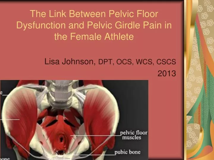
The Link Between Pelvic Floor Dysfunction and Pelvic Girdle Pain in the Female Athlete Lisa Johnson, DPT, OCS, WCS, CSCS 2013
Case: Pro Softball Player • Chief Complaints: • Constant, aching suprapubic and low back pain. • Intermittent left LE radicular pain to calf. • Intermittent vaginal pressure, severe cramping, painful defecation and increased urinary urgency. • Worse with running and jogging. • Pain Scale: 6/10 (rest) / 10/10 (activity)
History: • While catching a football pass four years prior, the pt. felt a sudden, sharp vaginal pain which persisted and progressed to the pubic and rectal regions. • Symptoms have progressively worsened. • PMH: • Long history of low back pain. • Treated by chiropractor (x 3yrs), PT, acupuncture and massage therapy with minimal benefit. • MRI: • (+) L 4-5 HNP
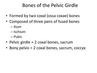
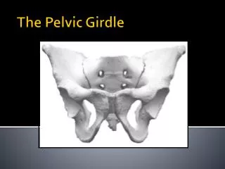
Pelvic Floor Anatomy The pelvic floor consists of five layers: III – Pelvic Diaphragm / Levator AniMuscles (Deep): 1. Pubococcygeus 2. Puborectalis 3. Iliococcygeus 4. Ischiococcygeus (Coccygeus)
Pelvic Floor Anatomy III – Levator Ani Muscles (Deep): • Pubococcygeus: • Arises from the dorsal surface of the pubic bone and obturator internus fascia, inserts on the anococcygeal and perineal bodies, anal wall. • Forms a hammock to support the urethra, vagina and rectum. • Pulls the rectum toward the pubic bone.
Pelvic Floor Anatomy III – Levator Ani Muscles (Deep): 2. Puborectalis: • Arises from the medio-lateral, dorsal surface of the pubic bone, blends with the lateral walls of the anus and rectum, and inserts at the external anal sphincter and anococcygeal body. • Controls descent of feces by elevating and constricting anal canal.
Pelvic Floor Anatomy III – Levator Ani Muscles (Deep): 3. Iliococcygeus: • Arises from the ischial spine and fascia of obturator internus, inserts to the anococcygeal body, anal wall and the coccyx. • Pulls the vagina and rectum toward the pubic bone. • Most widely recognized source of peri-anal referred pain to the sacrum, coccyx, rectum, vagina and lumbar spine.
Pelvic Floor Anatomy III – Levator Ani Muscles (Deep): 4. Ischiococcygeus (Coccygeus): • Originates on the ischial spine and inserts on the caudal aspect of the sacrum and the coccyx. • Provides tension to the pelvic floor, but not truly part of levator ani. • Pulls the coccyx forward and stabilizes the sacroiliac joint. • Innervated by ventral rami S4-S5
Pelvic Floor Function • Supportive: to the pelvic/abdominal organs. Elevates the pelvic floor, resisting increases in intra-abdominal pressure. • Sphincteric: Relaxes and contracts the urethral, vaginal and rectal openings. • Sexual: Maintains clitoral erection, provides tone and proprioception to the vaginal wall.
PELVIC FLOOR DYSFUNCTIONS • Two types of pelvic floor dysfunctions: • Hypertonus Dysfunctions (pain) - 15% of women have chronic pelvic pain. - Persistent or recurrent pelvic pain (> 3 mos) associated with symptoms of lower urinary tract, sexual, bowel or gynecological dysfunction. No proven infection or obvious pathology. - More common in women 26-30 (Steege, 1996) - Hypertonicity of the PFM often arises in young, very fit women with a hypertonic abdominals, preventing PFM relaxation.(Sapsford et al 2001)
PELVIC FLOOR DYSFUNCTIONS • Supportive Dysfunction (weakness) • Incontinence (UI) and Prolapse - Prevalence rates: 10-55% general population 28-49% HS/college athletes 52% elite athletes. (Thyssenet al, 2002) • Athletic activity can affect the development of (UI), depending on the extent of intra-abdom pressure and the strength of impact forces involved. (Bourcier et al. 1996) - Highest prevalence in sports involving high impact such as gymnastics, track and field and some ball games. (Bo, 2004)
PELVIC FLOOR HYPERTONUS DYSFUNCTIONS • Symptoms: Primarily PAIN! • Lumbar, perivaginal, perirectal, lower abdomen, coccygeal, posterior thigh. • Vulvar/clitoral burning • Dyspareunia (46% women-Steege, 1996) • Constipation • Common Diagnoses: • Vulvodynia, interstitial cystitis, levatoranisynd, coccydynia, pudendal neuralgia
PF Hypertonus Dysfunctions: Associated Myofascial Structures • Piriformis: • Can compromise pudendal nerve. • Refers pain into SI region, laterally to buttocks/posterior hip, 2/3 posterior thigh. • Obturator Internus: • Tendinous attachment with levator ani. • Refers pain into vagina, occasionally to posterior thigh, feeling of “fullness” of rectum.
PF Hypertonus Dysfunctions: Associated Myofascial Structures • Hip Adductors: • Adductor Magnus : • Refers pain deep into groin, pubis, vagina, rectum. • Usually “sharp, shooting” pain. • Pectineus: • Refers pain deep into groin, anterior hip joint, below inguinal ligament.
Case: Evaluation • Assess lumbar spine, SI joint, hips • Lower quarter muscle strength, tone and length • Lower quarter neuro screen • Assess pelvic floor muscles • External and internal digital exam • Sensation • Symmetry • Tone • Strength
Case: Objective Findings • Tenderness to palpation: (Severe) pubic symphysis, lower abdom, pirif, levator ani mus; (Mod) lumbar L3-5, sacrotuberous lig, obturator int, sacrococcygeal region. • ROM: Minimal limitation in trunk ext. • MMT: R LE – 5/5; L LE – 4/5 • Neural: ANTT L sciatic nerve, myotomal weakness L4-S1, Diminished L DTR • Structural: R/L Backward sacral torsion Left posterior innominate, lumbar rot. right
Physical Therapy Intervention • Manual Therapy & Therapeutic Ex • Joint mobilization – lumbar, SI jt, hip • Soft tissue mobilization • External – lumbar, pirif, OI, IP, abdom.CTM • Internal – pelvic floor muscles, OI • Exercise Program • Lumbar stabilization • Aerobic conditioning • LE muscle strengthening and flexibility • Modalities – ES, biofeedback to relax PF • Postural ed / body mechanics