Download

1 / 40
400 likes | 504 Views
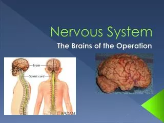
Homeostasis – Nervous System. Chapter 9. The Nervous System – 9.1. Vertebrate Nervous System is a highly organized communication network that allows the brain to gather information about the body, process that information and then act on it. Two main divisions:

E N D
Homeostasis – Nervous System Chapter 9
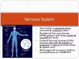
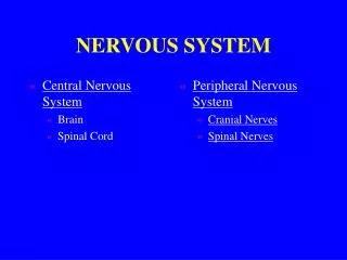
The Nervous System – 9.1 • Vertebrate Nervous System is a highly organized communication network that allows the brain to gather information about the body, process that information and then act on it. • Two main divisions: • Central Nervous System brain and spinal cord • Peripheral Nervous System nerves that carry information between organs and the CNS • Somatic Nerves skeletal muscle, bones, skin • Sensory somatic nerves relay information about environment to CNS • Motor somatic nerves initiate an appropriate response • Autonomic internal organ control • Sympathetic Nervous System nerve cells that prepare the body for stress. • Parasympathetic Nervous System nerve cells that return the body to a normal state after stress.
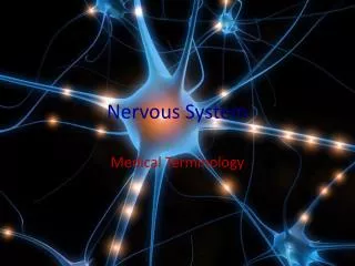
Anatomy of a Nerve Cell • Nervous system is made up of glial cells and neurons • Glial Cells non-conducting cells responsible for structural support and metabolism of nerve cells • Four main functions of glial cells: to surround neurons and hold them in place, to supply nutrients and oxygen to neurons, to insulate one neuron from another, and to destroy pathogens and remove dead neurons. • Neurons functional units of the nervous system • Sensory neurons sense and relay information from the environment to CNS; located in clusters called ganglia • Interneurons link neurons within the body, predominate brain and spinal cord • Motor neurons relay information to the effectors (muscles, organs, glands)
Anatomy of a Nerve Cell • Neurons contain: • Dendrites receive information and conduct information toward cell body • Cell bodies – main area of the neuron, contains nucleus • Axons – project information from the cell body; cytoplasmic extension of cell • Can be covered with a fatty protein called a myelin sheath that increase the rate of the impulse message. Formed by Schwann cells. Areas in between myelinated regions are called nodes of Ranvier.
Reattaching and Regenerating Nerves • Peripheral Nervous System does allow re-growth of nerves whereas the CNS does not. • Neurilemma surrounds the axon of PNS nerves and allows regeneration of severed neurons • Neurilemma and myelin sheath white matter of brain; if not grey matter which does not regenerate after damage • Nerve damage paralysis • Reattachment of nerves sometimes will facilitate re-growth with working connections • Grafting from peripheral nerve to the CNS • Stem cells • Spore like cells (3um big)
Neural Circuits • Reflex arc – simplest nerve pathway that allows you to react using a neural circuit through the spinal cord, not involving the brain. • Burn example
Electrochemical Impulse – 9.2 • Electricity in the Body • ECG electrocardiogram records the transmission of electrical impulses in heart muscle. • EEG electroencephalograph is used to measure brain-wave activity.
Electrochemical Impulse – 9.2 • Comparison of nerve impulse to electricity • Signal is faster along the wire, cytoplasmic core offers resistance • No diminishment of signal in nerve cells • Cellular energy creates currents not an external source.
Action Potential • Action Potential– the voltage difference across a nerve cell membrane when the nerve is excited. • Squid giant axon with electrodes inside and out, • nerve resting charge was -70mV, when excited the nerve had an electrical potential of +40mV. • Difference was only a few milliseconds then returned to -70mV (resting potential).
Action Potential • How does this happen? • Electrochemical event is caused by an unequal concentration of positive ions across the nerve cell membrane • Potassium ions inside the cell move out of the cell (via ion gates) and sodium ions move into the nerve cell. 50x more K+ moves out than Na+ in. • Outside becomes more positive so consequently inside becomes negative (-70mV is the resting potential of a polarized membrane. (-90mV would mean that more K+ ions had moved out of the membrane) • When excited the nerve cell become more permeable to Na+ than K+. This allows Na+ to rush into the nerve cell (depolarization)
Action Potential • A Na+/K+ pump restores the initial condition in the ratio of 3Na+ ions out of the nerve cell for every 2 K+ ions in. ATP runs this pump and this pump allows for a return to the resting potential through a process known as repolarization. • Depolarization & repolarization must occur before the process can happen again. The process of repolarization is called the refractory period
Action Potential • Step 1. Initiation • ► When a stimulus is encountered a closed voltage-gated Na+ channel is opened allowing Na+ ions to move into the neuron. • ► This causes the interior to develop a slight positive charge relative to exterior. • ► The Na+ voltage-gated channels close and the K+ voltage-gated channels open to assist the K+ ion channels in sweeping out positive charges to return the neuron to the resting potential. • ► This depolarization and restoration only takes about 5 milliseconds.
Action Potential • Step 2. Transmission • ► Transmission along a neuron is when the depolarization of one site stimulates the depolarization of adjacent sites. • ► The depolarization of one site is enough to cause nearby voltage-gated Na+ channels to open and permit Na+ ions to come into the neuron thus causing depolarization in an adjacent site. • ► The initial depolarization usually occurs at one end of a neuron and hence travels along the neuron in one direction however if the initial depolarization occurs in the middle, the action potential can travel in both directions. • ► A total jump of 110 millivolts of the inside of the neuron is required to send an action potential (-70 up to +30). • Refractory Period – the voltage-gated Na+ channels need to recover from their active state and while it only takes a fraction of a second, no action potentials can be transmitted.
Action Potential • Speedy Transmission • the speed of the action potential is determined by a few factors • one of those factors is the diameter of the axon. The larger the diameter, the more quickly the action potential will be sent • insulating the axon is another way to increase the speed of the action potential • Myelin sheath does not allow ion channels to open underneath them • Only at the node of Ranvier is the axon exposed so channels can open and close causing changes in polarization • The action potential then jumps from one node of Ranvier to the next thereby increasing the speed at which the action potential is sent. • The ion channels are concentrated in the nodes of Ranvier and this makes passing an action potential metabolically advantageous because there are fewer channels requiring ATP. • This is known as saltatory conduction
Movement of the Action Potential • Process above is only valid and will only elicit a response if the zone of depolarization is continued in adjacent regions. • A wave of depolarization is followed by a wave of repolarization.
Threshold Levels • Threshold level minimum level of a stimulus required to produce a response. • All or none response a nerve or muscle fibre responds completely or not at all to a stimulus. • Warm vs. Hot how do we tell the difference? • Frequency of impulses being sent to the brain. • Different neurons in an area having different thresholds.
Synaptic Transmission • Synapse – a small region between neurons or between neurons and effectors. • Neurotransmitters are chemicals contained in vesicles that get released from the presynaptic neuron and depolarize the postsynaptic neuron over a distance of about 20nm.
Synaptic Transmission • Slows the transmission down (not too many synapses in a reflex arc, but many involved in problem solving). • Acetylcholine – neurotransmitter that opens sodium ion channels causing depolarization broken down by enzyme cholinesterase • Excitatory (more permeable to Na+) or Inhibitory (more permeable to K+). Inhibitory means the neuron is said to be hyperpolarized (more negative). • Summation effect produced by the accumulation of neurotransmitters from two or more neurons.
The Central Nervous System – 9.3 • Brain and spinal cord • Meninges protective membrane that surround the brain and spinal cord • Three layers: dura mater (outer); arachnoid mater (middle); pia mater (inner) blood-brain barrier. • Cerebrospinal fluid cushioning fluid between the arachnoid and pia mater acts as a shock absorber and a transport medium • Lumbar puncture or spinal tap sample of CSF to diagnose bacterial or viral infection (i.e. meningitis)
The Spinal Cord • Sensory nerve messages from receptors brain motor nerve messages to mucles, organs and glands. • White matter myleinated nerve fibres from the sensory and motor neurons • Grey matter nonmyleinated interneurons • Made of many spinal nerves (when grouped is called a plexus)
The Brain • Brain complexity is what distinguishes humans from other animals • Poor in comparison agility, hearing, vision, smell • Good in comparison reasoning • Developmental links to other chordates forebrain, midbrain & hindbrain
The Brain • Forebrain • olfactory lobes (smell) • cerebrum (speech, reasoning, memory, personality). The surface of the cerebrum is known as the cerebral cortex which has many grey matter fissures for increased surface area. • Corpus callosum links one side to brain to the other as each side stores its own information…that is, one side is not the same as the other. • Hemispheres broken down into four parts; frontal, temporal; parietal; occipital • Thalamus located below cerebrum and below thalamus is the hypothalamus
The Brain • Associative Cortex – of the rest of the cerebral cortex a small portion is used by the motor and sensory cortex and the rest is used for thinking. In a mouse 95% of the cerebral cortex is used by sensory and motor areas whereas in humans only 5% is utilized for those functions with the rest being devoted to the associated cortex • Broca and Wernicke’s Areas • the left side of the brain is known to be associated with several speech centers. In right handed people B and W areas are located on the left side of the brain however in left handed people about half have these areas in the right side of their brain
Broca’s Area • controls the muscles necessary for speech. Injury to this area destroys the ability to speak altogether but not to read or write. • Wernicke’s Area • controls the form, or syntax, of speech injury to this area produces fluent, grammatical but meaningless speech
The Brain • Midbrain less developed than forebrain • Relay centre for some eye and ear reflexes • Hindbrain • Cerebellum located underneath the two cerebral hemispheres; controls limb movement, balance and muscle tone • Pons relay station between cerebellum and the medulla • Medulla oblongata major centre of autonomic nerve control (heart rate)
Brain Mapping • Phrenology – a pseudoscience that relates metal capacity to bumps on the head. Large brain means you are more intelligent. • Wilder Penfield – recognition and speech were in different areas…lead him to work toward mapping the function of different parts of the brain. • Interesting fact no sensory receptors in the brain
Neuroimaging • The key here is noninvasive why? • PET – positron-emission tomography reveals physiological and biochemical pathways • Radioactive isotope goes to different areas of the brain once tasks are to be completed and positrons interact with oppositely charged electrons resulting in radiation that is detected by a PET camera. • MRI – magnetic resonance imaging both as structure (MRI) and function (fMRI) • Nuclei of hydrogen atoms are usually random in order, and MRI uses magnets to align these protons/neutrons and radiowaves to knock these nuclei out of alignment. When knocked out of alignment faint radio signals are detected by an MRI scanner. • More water means more images produced which translates into a dark image. • CT – computerized tomography • Produces images of thin X-ray sections of body and can reassemble those images as a 3D image
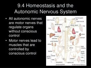
Homeostasis and the Autonomic Nervous System – 9.4 • The peripheral nervous system contains: • Autonomic nerves motor nerves that regulate organs of body without conscious control • Motor somatic nerves lead to muscles that are regulated by conscious control • Autonomic system (p.435, table 1) • Sympathetic Nervous System – prepares body for stress • Parasympathetic Nervous System – returns body to normal after stress • Vagus Nerve major cranial nerve that innervates the heart, bronchi, liver, pancreas, digestive tract
Painkillers • Endorphins (“endogenous morphine") and enkephalins natural painkillers produced in brain that counteract pain neurotransmitters in the substantiagelatinosa (SG) located in the spinal cord.
Painkillers • The substantiagelatinosa is one point (the nucleus proprius being the other) where first order neurons of the spinothalamic tract synapse. • Many μ- and к-opioid receptors, presynaptic and postsynaptic, are found on these nerve cells; they can be targeted to manage pain. • Opiates work the same way
Addictions • Physical, psychological and physiological • Physiological effects • structural changes will occur to neurons after continued consumption of certain drugs. These changes are not permanent however the symptoms of neuronal change can be witnessed when an individual goes through withdrawal symptoms.
Addictions • Neuromodulators can slow down the destruction of a neurotransmitter in the synaptic cleft so the neurotransmitter is present for a longer then required period and will continue to bind to receptors on the postsynaptic membrane. Other neuromodulators release neurotransmitters more easily from the presynaptic membrane or inhibit the reabsorption of the neurotransmitter.
Addictions • The action of drugs is that of a neuromodulator. They increase the amount of neurotransmitters in the synaptic cleft and thus the repeated binding of the neurotransmitter to the receptors in the postsynaptic membrane. What happens is that the receptors in the postsynaptic membrane reduce in number as there are too many transmissions being made.
Addictions • Without the drug the transmission now fails as not enough neurotransmitter is being released, and more importantly it is not staying in the synaptic cleft long enough to allow action potentials to be transmitted. This is why higher and higher levels of the drug need to be taken to obtain the same response as was achieve early in the addiction. • The number of receptors will be returned to about normal once the individual stops taking the drugs for a prolonged period of time. • Withdrawal symptoms, can be fatal