Download
1 / 23
230 likes | 341 Views
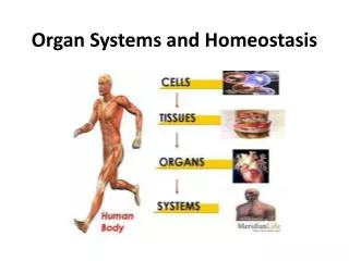
What are the other three primary tissue types? 9/25. Review the origins of the four primary tissue types What extracellular fibers are found in connective tissue? What is the function of connective tissue? What are characteristics of the ten different connective tissues?

E N D
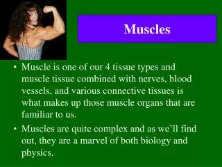
What are the other three primary tissue types? 9/25 • Review the origins of the four primary tissue types • What extracellular fibers are found in connective tissue? • What is the function of connective tissue? • What are characteristics of the ten different connective tissues? • What makes a cell “excitable”? • Neural tissues: supportive and nerve cells
Embryogenesis is the development of tissues and organs. This is a mini-review of embryonic development which creates ectoderm, mesoderm and endoderm.
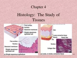
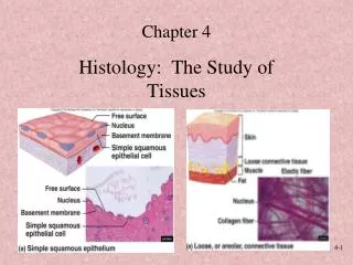
The Eight Kinds of Epithelial Tissue: Describe the key features of each and where they are found in the body: Four Single Layer Epithelial Tissues: • Simple Squamous Epithelial • Simple Cuboidal Epithelial • Simple Columnar Epithelial • Pseudostratified Columnar Epithelial Four Layered Epithelial Tissues: Stratified Squamous Epithelium-Keratinized • Stratified Squamous Epithelial-Nonkeratinized • Stratified Cuboidal Epithelial • Transitional Epithelial
Stratified Epithelial cells (Table 5.3) can be: 1) Squamous with keratin 2) Squamous without keratin, 3) Cuboidal or 4) Transitional. • Callus on Sole of Foot: SSE-K...creates a nasty water insoluble place where bacteria can’t live and water can’t evaporate from the body!…why? • Mucosa of vagina, mouth, anus, esophogus are covered by: SSE-NoK…why? • Ovarian follicles and seminiferous tubules of the testes: SCE…why? • Transitional Epithelium: Lower urinary tract and part of umbilical cord- it stretches! • Why transitional (partly rounded/partly flattened)? • WHAT IS EPITHELIAL CELL “EXFOLIATION”? • Do you treat the exfoliation on your scalp?
Connective tissues hold the body together, support the body, and transport materials within the body. • Connective Tissues are derived from Mesoderm: Functions: • Tendons vs. Ligaments- • Space filling materials- • Fat storage and insulation- • Blood cells and transportation- • How is connective tissue spacing different from that of epithelial cells? • What are the “extracellular” components of connective tissue?
What are the ten types of connective tissue? • Two Categories of CT called Fibrous or “fibroconnective” differ in fiber abundance (this makes up 5 CT types): • Dense CT: #1Dense Regular CT or #2Dense Irregular CT • Loose CT: #3Areolar CT #4Reticular CT #5Adipose • Cartilage has a rubbery matrix for support (3 CT types): “Chondrocytes in lacunae” • #6Hyalin Cartilage • #7Elastic Cartilage • #8Fibrocartilage • Richly calcified support: #9Bone • Osteocytes make up this CT • Fluid CT: #10 Blood and formed elements • Erythrocytes, Lymphocytes, Platelets
How do extracellular fibers contribute to the function of connective tissues? Fibroblasts: produce the fibers and ground substance in CT! • Collagen=25% of body protein/white • Stronger than steel by weight • Elastic Fibers: elastin/yellow • Where do we stretch? • What causes age lines in our skin? • Reticular Fibers: collagen+glycoproteins • Where is spongy framework? • Glycoprotein coated collagen • Why does Ground Substance fill space? How do G.S. contents promote the hydrogen bonding of water? • G.S. Contents: heparin, salts/minerals, glycosaminoglycan, chondroitin sulfate, heparin, hyaluronic acid of joints
Tendons consist of many collagen fibers. Collagen “fibers” are arranged in a staggered overlapping pattern of “fibrils”. This pattern imparts tremendous strength with almost NO stretch!
Rubbery elastin stretches and snaps back into the original shape! It is rich in negative charges that repel each other to help prevent “excess” stretching and to help it snap “back”.
Why is Blood one of the most important connective tissues? Can you live without blood? Can you live with too much blood? Cellular Components: • Anucleate red blood cells • Nucleated leukocytes for protection • “Leukemia” Critical Acellular Components: • Fibrinogen for clotting • Platelets • Plasma proteins/Clotting Factors • Lipoproteins • Immunoglobins • Nutrients/Waste • Plasmin for fibrinolysis
Areolar Tissues Underlies and attaches epithelia to underlying tissues! Contains scattered collagen, elastin and ground substance Protects vessels, nerves, trachea, visceral layers “Arena” for: immune defense, waste/nutrient transport Reticular tissues Tissues inside bone, lymph nodes, spleen and thymus Creates supportive framework to permit other cells to proliferate/reside Rich in “reticular” fibers and lymphocytes What are the characteristics of the three types of “loose connective tissue”? (Areolar, Reticular and Adipose)?
Dense Regular CT Fibroblasts: collagen/elastin fibers: “White” or “Yellow” Dense packing in parallel Super Strength in one stress angle! WHERE? Elastin sheets in blood vessels Tendons to muscle Ligaments between bones Dense packing leaves little space for blood vessels so injuries heal very slowly! Dense Irregular CT Fibroblasts: collagen/elastin fibers Dense packing in clusters Clusters in multiple angles Varied stress angle applied to DICT WHERE? Dermis of skin Protective organ capsules: kidney, bones, nerves, What two types of connective tissues contain large numbers of fibroblasts and dense collagen? How do they differ?
Where is Saladin’s typo in the connective tissues shown below? Which of these looks like a “Dense Regular” Connective Tissue? XXXXXXX XXXXXXX
What are the three different types of cartilage?All have cartilages have a covering called the perichondrium.What is the problem with Chondrocytes, Lacunae and Injury? • Hyalin Cartilage: dispersed collagen, chondriotin sulfate and a proteoglycan core • Your fetal skeleton is converted to bone later • Also: nose, trachea and bronchi. • Have you injured the costal cartilage of a rib? • Elastic cartilage: Dispersed collagen and elastin • Ear and Epiglottis • Flexible and elastic • Fibrous cartilage: Resists compression • Greater collagen content and density • Pubic symphysis, knee, vertebral discs
What does ground substance look like? The key is a large number of polar functional groups (negatively charged: -COO- or –SO4-) that attract water via H-bonds and repel other negatively charged ground substance if the other parts get too close. This creates a watery material that helps things slide across with minimal friction and lets it fill lots of space.
Composed of calcium matrix Collagen is critical! Two kinds of Osteocyte: Osteoblast-Makes bone Osteoclast-Erodes bone Also: Osteon, Lacunae, Lamellae, Canaliculi, Haversian Canal How is cell activity modified by aging and exercise? What limits the healing rate of broken bones? Spongy Bone has a hollow space filled with red or yellow marrow. Compact bone provides structure to support the body against gravity! When does this system fail us?All bones are wrapped in a fibrous sheath, the periosteum!
What is an “excitable” cell? (THE BASICS) Typical Excitable Cells: neurons and myocytes • All cells have a Na+/K+-ATPase that maintains a voltage across the membrane called a membrane “potential” • “Excitable” cells create a self-propagating wave of depolarization that moves across the plasma membrane. This wave represents information. • Depolarization can be used to promote changes in intracellular function: • Exocytosis and hormone/neurotransmitter release • Contraction • Changes in excitable cells may return to normal once the Na+/K+-ATPase has a chance to catch up
Nervous Tissues consist of excitable neurons and non-excitable supportive cells • Neurons: Can generate a self propagating action potential • Parts of a neuron: • Dendrite • Soma • Axon • Synapse • Why do neurons need supportive glial cells? • Fragility and excitable function • Glial cells are not excitable Health implications of glial cells for neural function?
Neural Tissues are derived from the primordial ectoderm (“skin like”). Remember that neural tissue is composed of two types of cell: neuron and glial
What do neural tissues look like?Neuron: Excitable vs. Glial Cell: Non-excitable
Synaptic transmission is the mechanism that permits a neuron to send its action potential to a second cell. How does it use vesicles to do this?