Download
1 / 21
210 likes | 565 Views
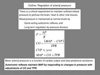
Outline: Regulation of arterial pressure. There is a critical requirement to maintain sufficient blood pressure to perfuse the brain, heart & other vital tissues. Blood pressure is maintained at normal levels by Quick-acting autonomic reflexes and Long term regulation by pressure diuresis.

E N D
Outline: Regulation of arterial pressure • There is a critical requirement to maintain sufficient blood pressure to perfuse the brain, heart & other vital tissues. • Blood pressure is maintained at normal levels by • Quick-acting autonomic reflexes and • Long term regulation by pressure diuresis Mean arterial pressure is a function of cardiac output and total peripheral resistance. Autonomic reflexes maintain MAP by responding to changes in pressure with adjustments of CO and TPR.
Carotid sinus nerve to glossopharyngeal n. (IX) Vagus n. (X) Carotid sinus Carotid body Common carotids Aortic bodies Aortic arch Part 1: Quick acting Autonomic Reflexes that maintain arterial pressure The arterial baroreflex is the most important autonomic reflex maintaining MAP. This reflex can induce changes CO & TPR within seconds in response to a change in MAP. Input • SENSORS • Baroreceptors • Carotid sinus • Aortic arch • Atria • Vena cava • Chemoreceptors • Peripheral • Aortic body • Carotid body CNS (set point) Output Sympathetic Parasympathetic hormonal Feedback change
CNS Glossopharyngeal Vagus Hering’s nerve Aortic arch Carotid sinus 80 100 120 140 160 180 Carotid sinus pressure response Impulses/sec carotid sinus nerve Arterial pressure, mm Hg The baroreflex senses changes in arterial pressure as changes in diameter of the carotid sinus & aortic arch. An increase in diameter stretches mechanoreceptors in the walls of the vessels. The frequency of action potentials from the receptors is directly proportional to arterial pressure.
Cerebral cortex Excitatory or inhibitory Anterior hypothalamus Posterior hypothalamus Vasomotor center (Medulla & Pons) Excitatory or inhibitory Excitatory A2 Sensory area (nucleus tractus solitarius) Vagus & glossopharyngeal nerves Vascular baroreceptors Central input to vasomotor area Afferent limb of the baroreflex Arterial input to vasomotor area
Vasomotor center (Medulla & Pons) Vasoconstrictor area C 1 vasoconstriction sympathetic Efferent signals from the vasomotor center Dorsal motor nucleus Vasodilator area A1 inhibition Vagus nerve Sino-atrial node (heart rate) parasympathetic Almost all small arteries, arterioles, venules & veins have sympathetic constrictor innervation. Changes in sympathetic activity can affect TPR. Sympathetic nerves also carry vasodilator fibers to skeletal muscle.
+ blood volume Arterial blood pressure Baroreflex Venous mechanoreceptors (atria, vena cava near heart) arterial mechanoreceptors (carotid sinus,aortic arch) IX, X secretion of NaCl & H2O retaining hormones Central nervous system sympathetic tone Parasympathetic tone to heart venous pressure contractility HR venous return stroke volume CO = HR x SV cardiac output TPR MAP = CO x TPR Arterial blood pressure
Pressure range of baroreceptors Carotid sinus afferents are most important in regulating arterial pressure in the normal pressure range. Chemoreceptors primarily regulate blood pH, PCO2 and PO2; at low arterial pressure they potentiate vasoconstriction & stimulate respiration. Increased respiratory rate aids in venous return.
CNS ischemic response Blood flow to brain Blood flow to vasomotor center PCO2 in vasomotor center Maximal stimulation of sympathetic outflow • The CNS ischemic response is a “last-ditch” emergency response that produces maximal increases in arterial pressure (up to 250 mm Hg). • The CNS ischemic response produces its maximal effect when arterial pressure is in the 15 to 20 mm Hg range.
Venous blood reservoirs & baroreflex The capacitance veins in the liver, lungs, spleen, intestines & subcutaneous venous plexus constrict with sympathetic stimulation as part of the baroreflex response to hypotension or hypovolemia. Veins feeding into the superior vena cava do not participate in baroreflex induced constriction. Constriction of the capacitance veins transfers blood toward the heart. Therefore these veins act as blood reservoirs.
150 125 100 Arterial pressure, mm Hg 75 50 25 0 5 10 15 20 25 Continuous vasoconstrictor tone from the vasomotor center maintains arterial pressure. Norepinephrine I.V. Sympathetic block Sympathetic blockade decreases AP by ~ 50% Minutes • Spinal block via injection of anesthetic blocks sympathetic tone from medulla • Arterial pressure decreases ~ 50% • Norepinephrine can still elicit a constriction (vessels are responsive) • Quadriplegia • Arterial pressure is unstable, heart rate decreases, stroke volume increases
Cutting the baroreflex nerves makes the arterial blood pressure unstable. control no baroreflex
Emotional disturbance (cerebral cortex) Anterior hypothalamus Adrenal medulla Vagus nerve medulla Spinal cord b receptors Heart rate Vasodilator fibers to skeletal muscle epinephrine cardiac output Dilation in skeletal muscle TPR arterial pressure Brain blood flow Fainting (syncope) Vasovagal syncope
Healthy young Healthy old Autonomic failure 160 140 MAP before test meal 120 MAP, mm Hg 100 80 D MAP = -25 mm Hg 60 40 20 0 30 60 90 0 30 60 90 0 30 60 90 Minutes after a standard test meal Postprandial hypotension in autonomic insufficiency
Circulatory changes in the postprandial state in healthy young subjects Ingestion of a meal Afferents from GI tract CNS Sympathetic activity Gut metabolism Gut hormones Sympathetic activity Resting resistance skeletal muscle, skin Resistance in GI tract TPR maintained at normal level Maintenance of MAP via interaction of local metabolic effects with changes in sympathetic tone is a general principle of circulatory function.
Ingestion of a meal Afferents from GI tract CNS Gut metabolism Gut hormones Sympathetic activity resistance skeletal muscle resistance in GI tract TPR decreases, MAP decreases Circulatory changes in the postprandial state in autonomic failure Subjects with autonomic failure experience a large decrease in MAP after a meal that may be accompanied by syncopedue primarily to absence of an increase in resistance in resting skeletal muscle, skin.
Use of the tilt table aids in determining the cause of syncope. Passive head-up tilt causes maximal dependent pooling of blood and stresses the baroreflex response to decreased central blood volume. Normal In autonomic failure hypotension causes syncope because the decrease in AP is not accompanied by the normal baroreflex mediated increase in HR. The two most common causes of postural hypotension are autonomic failure (which can be caused by multiple disorders) and volume depletion.
The pressure diuresis mechanism acts as a negative feedback connecting kidney function with blood pressure Arterial pressure Arterial pressure - Na+ excretion Na+ excretion + Urine flow Urine flow Blood volume Blood volume Diuresis: urine flow Natriuresis: Na+ excretion Part 2: Long term regulation by pressure diuresis
5 4 Na+ excretion 3 2 1 100 120 80 Intrinsic pressure diuresis in an isolated artificially perfused kidney In an isolated kidney only intrinsic mechanisms function; nerves have been severed and circulating hormones are not present Perfusion pressure pressure
Renal function curve 5 4 Na+ excretion 3 2 Intrinsic pressure diuresis curve 1 80 100 120 Arterial blood pressure Pressure diuresis versus renal function curve The renal function curves shows the effect of arterial pressure on Na+ excretion in the intact kidney with normal neural and hormonal function
normal Hypertension is a renal disease 5 Atrial natriuretic peptide (ANP) 4 Na+ excretion (times normal) 3 angiotensin II 2 1 Arterial blood pressure ANP Na+ excretion angiotensin II Na+ excretion 80 100 120 Hormones that affect renal Na+ reabsorption shift the pressure diuresis curve Angiotensin stimulates Na+ reabsorption, opposing pressure diuresis. Under angiotensin stimulation a higher blood pressure is needed to excrete the ingested NaCl. ANP has the opposite effect.
Short term Short and long-term control of arterial pressure Thirst Aldosterone Vasopressin CNS vasomotor center Long term Fluid intake Sympathetic activity Blood volume Fluid output (urine) Contractility Parasympathetic activity Preload Frank-Starling mechanism HR SV TPR CO Pressure diuresis MAP Baroreceptors