Arthritis
Arthritis. Clinical Quiz. DMARDs. Which drugs and how do they work?. Methotrexate. Folic acid antagonist → Cytotoxic, immunosuppressant. Sulfasalazine. Scavenger of toxic metabolites produced by neutrophils. Penicillamine. Immunosuppressant. Blocks IL-1 and collagen maturation. Gold.


Arthritis
E N D
Presentation Transcript
Arthritis Clinical Quiz
DMARDs Which drugs and how do they work? Methotrexate Folic acid antagonist → Cytotoxic, immunosuppressant Sulfasalazine Scavenger of toxic metabolites produced by neutrophils Penicillamine Immunosuppressant. Blocks IL-1 and collagen maturation Gold Blocks IL-1, TNF Chloroquine Antimalarial
“Biologicals” What are the targets, and what effect rheumatic disease? Adalimumab Infliximab TNF → Anti inflammatory Entanercept Rituximab CD20 → Anti B Cell
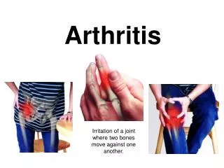
Rheumatoid Arthritis Clinical diagnosis requires ≥ 4 of these 7: • Morning stiffness > 1 hr for ≥ 6 weeks • Arthritis in ≥3 joints for ≥ 6 weeks • Arthritis of hand or wrist joints ≥ 6 weeks • Symmetrical arthritis • Rheumatoid nodules • Serum RhF • Typical radiological changes
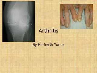
Imaging OSTEOARTHRITIS Standing radiograph of the knee reveals narrowing of the medial and lateral femorotibial compartments with marginal osteophytes.
Imaging RHEUMATOID ARTHRITIS Multiple erosions with deformity of the carpal bones in a patient with rheumatoid arthritis of the hands.
Imaging ANKYLOSING SPONDYLITIS Hip joint changes. Frontal radiograph shows uniform joint space narrowing and a collar of osteophytes at the femoral head-neck junction. Left sacroiliac joint ankylosis is present.