Download
1 / 13
220 likes | 864 Views
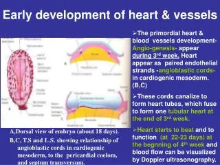
Early development of heart & vessels. The primordial heart & blood vessels development- Angio-genesis- appear during 3 rd week. Heart appear as paired endothelial strands - angioblastic cords- in cardiogenic mesoderm. (B,C)

E N D
Early development of heart & vessels • The primordial heart & blood vessels development- Angio-genesis- appear during 3rd week. Heart appear as paired endothelial strands -angioblastic cords- in cardiogenic mesoderm. (B,C) • These cords canalize to form heart tubes, which fuse to form one tubular heart atthe end of 3rd week. • Heart starts to beat and to function (at 22-23 days) atthe begnning of 4th week and blood flow can be visualized by Doppler ultrasonography. A,Dorsal view of embryo (about 18 days). B,C, T.S and L.S. showing relationship of angioblastic cords in cardiogenic mesoderm, to the pericardial coelom, and septum transversum.
Drawing of embryonic cardiovascular system(26 days), at 4th week showing left vessels : • Umbilical vein. • Vitelline vein. • Cardinal veins : anterior, posterior,& common cardinal v.
Development of veins associated with the heart of 4th week embryo • Vitelline veins return poorly oxygenated blood from yolk sac to the sinus venosus of heart. • Umbilical veins carry well-oxygenated blood from primordial placenta to sinus venosus. • Common cardinal veins carry poorly oxygenated blood from body of embryo to sinus venosus
Dorsal views of the developing heart. • A, during 4th W. showing primordial atrium & sinus venosus and veins draining into them. • B, 7th W. showing venous circulation through liver. • C, 8th W,adult derivatives of anterior cardinal veins.
Vitelline Veins • Lie in the yolk stalk, they carry poorly oxygenated blood from yolk sac to sinus venosus passing through septum transversum. • In the region of developing liver in septum transversum, hepatic sinusoids develop fromvitelline veins, and Hepatic veins develop from remains of right vetilline vein. • Proximal left vitelline veindegenerates, but / proximal part ofright vitelline vein form hepatic part ofI.V.C. • Portal vein develops from an anastomotic network of distal partsof right & left vitellineveins around the duodenum.
Umbilical Veins • They carry well-oxygenate blood from placenta to sinus venosus. • As the liver develops, umbilical veins lose their connection with heart. • Right umbilical veindisappearscompletely. • Proximal (cranial) part of left umbilical vein between liver & sinus venosus degenerates. • Distal (caudal) part of left umbilical veinpersist and becomes the umbilical vein, which carries blood from placenta to embryo. How? • A large venous shunt- ductus venosus- develops within liver and connects umbilicalvein with I.V.C. so blood pass directly from placenta to heart.
Anterior Cardinal Veins • Cardinal veins constitute the main venousdrainage of the embryo. • Anterior & posterior cardinal veins drain cranial & caudal parts of embryo ,respectively. • A,4th week, anterior & posterior cardinal veins join the common cardinal veins, which enter sinus venosus. • Anterior cardinal veins are connected by anastomosis, which shunts blood from left to right. B,7th week. • This anastomotic shunt between anterior cardinal veins becomes left brachiocephalic vein when caudal part of left anterior cardinal vein degenerates. C, 8th week. • S.V.C. develops from right anteriorcardinal vein + right commoncardinal vein.C, 8th week.
Posterior Cardinal Veins • Posterior cardinal veins develop primarily as vessels of mesonephroi and disappear with transitory kidneys. • The only adult derivatives of posterior cardinal veins are : root of azygos vein + common iliac veins. Ventral views of primordial veins of the embryos’s trunk. A, 6 weeks. D, adult.
Subcardinal & Supracardinal Veins • Subcardinal veins appear first, followed by supracardinalveins to replace gradually the posterior cardinal veins. • Subcardinal veins are connected together through subcardinal anastomosis and with posterior cardinal veins through anastomosis throughmesonephros (early kidney),Also sub-supracardinalanastomosis develops. • These anastomosis shunt blood from left to right veins, as a result, right veins enlarge while left veins become small and may disappear. • Drawings illustrating ventral views of primordial veins of trunk : vitelline ,umbilical & cardinal veins, and also subcardinal & supracardinal veins. • A, 6 weeks. • B, 7 weeks.
Fate of Subcardinal veins • Left subcardinal veincranial to the anastomosisdisappears leaving small left suprarenalvein,while caudal toanastomosis it becomes left gonadal vein. • Right subcardinal veincranial to the anastomosis forms the pre-renal part of I.V.C. + right suprarenal vein,while caudal to the anastomosis it develops into right gonadal vein. • Sub-supracardinal anastomosis forms right & left renal veins + renal part ofI.V.C. • Ventral views of primordial veins of trunk’s embryo. • C, 8th week. • D, adult.
Fate of Supracardinal veins • They are the last pair of vessels to develop. • The middle part of the 2 veins in the region of kidney disappears. • Cranial part of left supracardinal vein + transverse anastomosis form Hemiazygos vein. • Cranial parts of right supracardinal vein + right posterior cardinal vein form Azygos vein. • Caudal to the level of kidney : right supracardinal vein forms postrenal part ofI.V.C., while left supracardinal veindisappears. • Ventral views of primordial veins of trunk’s embryo. • C, 8th week. • D, adult.
Development of I.V.C • Hepatic part : develops from hepatic vein (from proximal part of right vitelline vein) + hepatic sinusoids. • Prerenal part : develops from rightsubcardinal vein. • Renal part : develops from subcardinal-supracardinal anastomosis. • Postrenal part : develops from right supracardinal vein. .