Download
1 / 25
250 likes | 466 Views
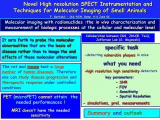
Novel High resolution SPECT Instrumentation and Techniques for Molecular Imaging of Small Animals F. Garibaldi - ISS-NIH, Rome, 4-6 June 06. Molecular imaging with radionuclides :the in vivo characterization and measurement of biologic processes at the cellular and molecular level.

E N D
Novel High resolution SPECT Instrumentation and Techniques for Molecular Imaging of Small Animals F. Garibaldi - ISS-NIH, Rome, 4-6 June 06 Molecular imaging with radionuclides :the in vivo characterization and measurement of biologic processes at the cellular and molecular level Collaboration between ISS, JHU(B. Tsui), Jefferson Lab (S. Majewski) It sets forth to probe the molecular abnormalities that are the basis of diseaserather than to image the end effects of these molecular alterations specific task -detecting vulnerable plaques in mice what you need -high resolution high sensitivity detectors key parameters: - SNR - FOV - Sensitivity - Spatial Resolution - simulations, prel. measurements The rat and mousehost a large number of human diseases. Therefore one can study disease progression and therapeutic response under controlled conditions PET (microPET) cannot attain the needed performances ! MRIdoesn’t have the needed sensitivity Summary and outlook
Ultrasound CT Optical (Bioluminescence, fluorescence) A F Structure A M 0.1 mm Unique !! Doppler Topography µm to mm ~103 cells quantitative Tissue Density, Z A 20-50 µm MRI PET/SPECT F M A F M Radiotracer ~1-2 mm H Concentration <10-12 mole = quantitative 0.1 mm BOLD, DCE Molecular Imaging Modalities -galactocidase 0.1 µmole H / µmole 31P
Patient injected with radioactive drug. Drug localizes according to its metabolic properties. Gamma rays, emitted by radioactive decay, that exit the patient are imaged. γ Imaging: Single Photon Detector Module • Collimator Only gammas that are perpendicular to imaging plane reach the detector • Readout Electronics Amplify electrical signal and interface to computer • Scintillator Convert gammas to visible light • Computer decoding procedure Elaborate signal and gives image output • Photomultiplier Convert light to electrical signal
High ResolutionHigh SensitivityDetectors FWHM = 7.4 mm Diffusive Wall FWHM = 5.4 mm Absorbing Wall key parameters SNR (and contrast) (spatial resolution) FOV S = counts in ROI, BKG = background Max = max. counts in tumor ROI they are correlated Gamma Emission X position Light Spread Function (LSF) energy resolution plays only a secondary additional role in imaging breast under compression
Important parameters fordetectability/visibility efficiency detector and collimation • SNR • - Contrast time (and modality) uptake (radiopharmacy) scintillator spatial resolution detector intrinsic properties pixel dim/n.of pixels modality (compression) electronics, DAQ . Uniformity of p.h.response (affecs the overall en res. and the energy window sel.) CsI(Tl) Bialkali PMT Bialkali PMT fotofraction
Importance of pixel identification good pixel identification is fundamental for correct digitization affecting spatial resolution and contrast C8 strips M16 (4 x 4) mm2 M64 (2 x 2) mm2
Projective coordinate electronics 1024 ch, 2 KHz Under study 4096 Ch. -> 8192 Ch. (10-20 kHz)
tumors: (5, 6, 7, 8,9,10,12) uptake 1:10; breast 6 cm NaI(Tl) 1.5 pitch; H8500 (6x6 mm2) measurements 6 mm 8 mm 7 mm X (H9500) NaI(Tl) 1.3 pitch; H8500(6x6 mm2) 6 mm tumors visible NaI(Tl) 1.2 pitch H9500 (3x3 mm2) measurements confirm simulation smaller scintillator pixel, higher SNR but tum 8 mm anode pixel has to be small
Performances not good enoughfor imaging biological process in vivo in small animals (mice) man rat Trying to Image apoptosis by proper tracer (e.g.99mTcINIC-Annexin-V) Geant 4 simulation detector area: 100 x 100 mm2 aorta: ~ 2 mm diameter plaque size: 0.5 x 1 x 4 mm3 • - pixellated CsI(Tl) (0.8 - 0.4 mm pitch) • LaBr3 continuous (3 mm thick, different surface(s) treatment(diffusive vs absorptive) • 6 x 6 mm2, 3 x 3 mm2, 1.5 x 1.5 mm2 • (PMT anode pixel size) spatial resolution: ~ 500 m system sensitivity: ~ 10 cps/Ci 1000 counts/view/resol.elem. 1 plaque = 10 mCi,10resol.elem.
Summary of CsI(Tl), pixellated 0.6 mm pitch H9500 0.6 mm pitch Burle What about CsI(Na) ?? LaBr3 to be carefully evaluated
Scaling down (100 mm, 0.8 mm --> 50 mm, 0.4 mm) 1/4 of detector area, 1/4 number of channels but 0.4 mm is very small !! mouse doesn’t scale !
simulation summary snr calculation sensitivity too small but multiply by ~ 4 (multipinhole) x 4(8) ( n. of modules) snr = 30 ( 60 ) ===> plaque “visible”
There are decoding patterns G allowing: A G = d then A G = Ô, in fact sensitivity smaller than required Coded apertures Ô = R G = ( O× A ) G = O * (A G) = O * PSF Submillimeter spatial resolution(FWHM=0.93 mm) AND High sensitivity (~850cps/MBq) 30 time pinhole already obtained recostruction possible in a deepth of focus as largeas large as 4 cm !! Both spatial resolution and sensitivity still to be improved F. Cusanno et al. NIM A Smaller scintillator pixels (0.8 --> 0.6 mm) ==> smaller photodetector anode pixels
Measurements 60Co source, 122 keV CsI(Tl) 1.0 pitch H9500 (6 x 6mm2) CsI(Tl) 1.25 pitch H9500 (3 x 3mm2) NaI(Tl) 1.25 pitch H8500 (6 x 6 mm2) measurements confirm simulations: small anode pixel is needed for small scintillator pixel (0.8 --> 0.6 mm --> high number of channels 1024 for 1 module! )
LaBr3 continuous 1.5 mm thick + H9500 (3 x 3 mm2 anode); 3 mm thick + H8500(6 x 6 mm2) Image of a 0.25mm slit. Chipped edge seen at left. Non-uniformities to correct. 1.5mm thick LaBr3 attached to a H9500 PSPMT Measured energy resolution ~ 8% FWHM @122 keV Projection of the image of the slit. FWHM = 0.65mm Mcarlo FWHM = 0.615 3 mm mm thick LaBr3 attached to a H8500 PSPMT Active area 0.75 mm FWHM Mcarlo FWHM = 0.8 Dead area 1.4 mm FWHM
Preliminary pinhole SPECT reconstruction results from the CsI detector Images are displayed as MIP (maximum-intensity-re-projections) animations Please use slide show mode to see the animation 2 point sources APOE mouse (kidneys shown) Sample projection image Flood image
Conclusions A solution for this challenging problem exists: - good results with CsI(Tl) 1 mm pitch + H9500 100 pixels, spatial resolution 0.53 mm - 0.46 mm, FOV=33-25, (22-39) cps/MBq • improvements needed? • scaling down (50 x 50 mm2, 0.4 mm pitch, Burle photodetector (3x3 mm2) ->more compact, much less expensive - 100 x 100 mm2 CsI(Tl) 0.8 (0.6) mm pitch with individual readout • careful evaluation of LaBr3 option (the advantage is better energy resolution • (very important if multilabeling shows to be possible and useful) • Fov, surface treatment, thickness, availability, cost • decisionto be taken on the base of SNR obtained with measurements (phantoms) Measurements for CsI(Na) 0.4 - 0.6 mm pitch, LaBr3 3 mm thick 10 x 10 cm2 vs 5 x5 cm2 (scaling down) (tomographic reconstruction will be decisive) Final layout on two steps next two years (if funding allows)
Invited talks Invited talks a Congressi Internazionali 1.F. Cusanno. “High Resolution, High Sensitivity detectors for Molecular imaging with Radionuclides: the Coded Aperture option”. Milos (Grecia). Imaging technologies in Biomedical Sciences. September 2005 2. F. Garibaldi. “High Resolution, High Sensitivity Detectors”, Advanced Molecular Imaging Techniques in the Detection, Diagnosis, Therapy, and Follow-Up of Prostate Cancer, Rome, 6-7 December 3. Magliozzi ML et al “High Resolution, High Sensitivity Detectors for Molecular Imaging of Small Animals and Tumor Detection”. International Conference of Advanced Detectors. Como (Italy), October17-21, 2005 4. F. Garibaldi, “Molecular imaging: high resolution detectors for early diagnosis and therapy of breast cancer”. Milos (Grecia). Imaging technologies in Biomedical Sciences. September 2005 5. "Molecular Breast Imaging: first results from Italian National Health Institute clinical trials", to be presented at the International Conference "Fist European Conference on Molecular Imaging Technology (EUROMEDIM2006)” Marseille, France, 9 - 12 May 2006 6. E. Cisbani, “Imaging with radionuclides: a powerful means for studying biological processes in vivo", Fist European Conference on Molecular Imaging Technology (EUROMEDIM2006)", Marseille, France, 9 - 12 May 2006 - Cuba ?
Publications • 1. F. Garibaldi et al. “A PET scanner employing CsI films as photocathode”, Nucl. Instr. Meth., 2004, A525, 263-267. • F. Garibaldi et al. “Novel design of a parallax freeCompton enhanced PET scanner”, Nucl. Instr. Meth., 2004, A525, 268-274. • 3. R. Pani, M.N. Cinti, F. Cusanno, F. Garibaldi et al“Imaging detector designs based on Flat panel PMT”, Nucl. Instr. Meth., 2004, A527, 54-57. • F. Cusanno et al. “Molecular imaging by single-photonemission”, Nucl. Instr. Meth., 2004, A527, 140-144. • F. Cusanno et al. “Preliminary Evaluation of Compact Detectors for Hand-Held Gamma Cameras”, Physica Medica, 2004, XX (2), 65 • Pani R. Cinti, M.N., Cisbani, E.; Colilli, S.; Cusanno, F.; De Vincentis 6. Preliminary study of metabolic radiotherapy with 188-Re via small animal imaging, A. Antoccia, G. Baldazzi, M. Bello, D. Bernardini, P. Boccaccio, D. Bollini, F. de Notaristefani, F. Garibaldi, G. Hull, U. Mazzi, G. Moschini, A. Muciaccio, F.-L. Navarria, V. Orsolini Cencelli, G. Pancaldi, R. Pani, A. Perrotta, M. Riondato, A. Rosato, A. Sgura, C. Tanzarella, N. Uzunov, M. Zuffa Nuclear Physics B, Volume 150, January 2006, Pages 411-416 • Small animal imaging by single photon emission using pinhole and coded aperture collimation, Garibaldi, F.; Accorsi, G.; Fortuna, A.; Fratoni, R.; Girolami, B.; Ghio, F.; Giuliani, F.; Gricia, M.; Lanza, R.; Loizzo, A.; Loizzo, S.; Lucentini, M.; Majewski, S.; Santavenere, F.; Pani, R.; Pellegrini, R.; Signore, A.; Scopinaro, F.; Veneroni, P.; IEEE Transaction on Nuclear Science, Volume 52, Issue 3, Part 1, June 2005 Page(s):573 – 579 • New Devices for Imaging in Nuclear Medicine, Cancer Biotherapy & Radiopharmaceuticals, 19(1), 121-128, 2004 • A PET scanner employing CsI films as photocathode, Nucl Instr Meth A525, 2004, 263-267 • 10. Novel design of a parallax free Compton enhanced PET scanner Nucl Instr Meth A525, 2004, 268-274
11. A study of intrinsic Crystal-pixel light-output spread for discrete scintigraphic imagers modeling, Scafe, R.; Pellegrini, R.; Soluri, A.; Montani, L.; Tati, A.; Cinti, M.N.; Cusanno, F.; Trotta, G,Pan Pani, R.;Garibaldi,F.,IEEE Transaction on NuclearScience, Volume 51, Issue 1, Part 1, Feb. 2004 Page(s):80 - 84 12. Custom breast phantom for an accurate tumor SNR analysis, Cinti, M.N.; Pani, R.; Garibaldi, F.; Pellegrini, R.; Betti, M.; Lanconelli, N.; Riccardi, A.; Campanini, R.; Zavattini, G.; Di Domenico, G.; Del Guerra, A.; Belcari, N.; Bencivelli, W.; Motta, A.; Vaiano, A.; Weinberg, I.N.; IEEE Transaction on Nuclear Science, Volume 51, Issue 1, Part 1, Feb. 2004 Page(s):198 - 204
Publications • - Molecular imaging by single-photon emission”, Nucl. Instr. Meth., 2004, A527, 140-144. • - Preliminary Evaluation of Compact Detectors for Hand-Held Gamma Cameras”, Physica Medica, 2004, XX (2), 65-70 • - Small Animal Imaging by Single Photon Emission Using Pinhole and Coded Aperture Collimation”, IEEE Tran Nucl Sci, 2005, 52(3), 573-579. • - Tumor SNR Analysis in Scintimammography by Dedicated High Contrast Imager”, IEEE Trans Nucl Sci, 2003, 50(5), 1618-1623 • Custom breast phantom for an accurate SNR analysis, IEEE Trans. N.S., Vol 51, N.1 Feb. 2004 • - Molecular imaging: high resolution detectors for early diagnosis and therapy monitoring of breast cancer, • To be published on NIM, Milos • High Resolution, High Sensitivity Detectors for Molecular Imaging with Radionuclides: the Coded Aperture Option, to be published on NIM • Euromedim Francesco • Euromedim Evaristo • Euromedim Carrato • A. Dragone
1. “A PET scanner employing CsI films as photocathode”, Nucl. Instr. Meth., 2004, A525, 263-267. • 2. “Novel design of a parallax free Compton enhanced PET scanner”, Nucl. Instr. Meth., 2004, A525, 268-274. • 3. “Imaging detector designs based on Flat panel PMT”, Nucl. Instr. Meth., 2004, A527, 54-57. • 4. “Molecular imaging by single-photon emission”, Nucl. Instr. Meth., 2004, A527, 140-144. • 5. “Preliminary Evaluation of Compact Detectors for Hand-Held Gamma Cameras”, Physica Medica, 2004, XX (2), 65-70. • 6. “Small Animal Imaging by Single Photon Emission Using Pinhole and Coded Aperture Collimation”, IEEE Tran Nucl Sci, 2005, 52(3), 573-579. • 7. Milos code apertures • 8. Milos breast? • 9. Prostate Rome, in preparation • 10. Invited talks at Euromedim (titoli anche se non so se mettere il breast) • 11. Deleo et al (la PET del CERN sottomesso a NIM) • 12. altri lavori (recenti) di PET CERN) • 13. lavori dei nostri amici del pin diodes (IEEE CD?? ) • 14. altra roba recente di Pani in cui ha messo solo me? • 15. Como M.Lucia • Invited talsk (presentazione a congressi): • 1. Milos 2005 • 2. Milos 2005 • 3. Prostate Conference • 4. Euromedim a Maggio • 5. What else? (dal 2004)? • 6. Como MLuci • 7. Cuba?
Positron Emission Tomography microPET
Detection coincident events between two detectors • Compton scatter equation relates scatter angle and Eo and Ere • Photon direction is determined within conical ambiguity Internal Compton Probe