Download
1 / 13
140 likes | 678 Views
Restriction Enzyme Cleavage of DNA . Carolina Kit. Timeline. Week Before: HW—DNA scissors, DNA goes to the races Monday—pour gels Tuesday—Lecture, Run gels with DNA, photograph gel Monday—Lab write-up due. Write-up. Annotate handout

E N D
Restriction Enzyme Cleavage of DNA Carolina Kit
Timeline Week Before: HW—DNA scissors, DNA goes to the races Monday—pour gels Tuesday—Lecture, Run gels with DNA, photograph gel Monday—Lab write-up due
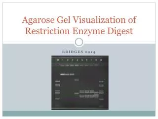
Write-up • Annotate handout • Data draw a gel and mark each banding site, staple picture to lab that you turn in to me • Results and Discussion (do the graph!)—answer all part
Background Information • Use website http://bioinformatics.dnalc.org/gmo/animation/gmo.html • Animations How did these samples get cut? Cutting and pasting A & B Why can restriction enzymes be used for? Transferring and storing A & B
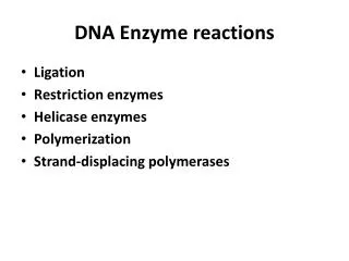
Important for this lab: • Endonucleases – enzymes that cut RNA or DNA at specific sites; restriction enzymes are endonucleases that cut DNA • Sticky cells – restriction fragments in which one end of the double stranded DNA is longer than the other; necessary for the formation of recombinant DNA • Restriction enzyme mapping – determining the order of restriction sites of enzymes in relation to each other • Restriction enzymes are used for transformation (we will do this soon): • Transformation – the uptake and expression of foreign DNA by a cell • Transduction – the use of viruses to transform or genetically engineer cells • Competent/competency – the ability of cells to take up DNA • Selection – the process of screening potential clones for the expression of a particular gene, for example, the expression of a resistance gene (such as resistance to ampicillin) in transformed cells • Transformation efficiency – a measure of how well cells are transformed to a new phenotype • Recovery period – the period following transformation where cells are given nutrients and allowed to repair their membranes and express the “selection gene(s)” • Beta-galactosidase gene – a gene that produces beta-galactosidase, an enzyme that converts the carbohydrate X-gal into a blue product • Green fluorescent protein – a protein found in certain species of jellyfish that glows green when excited by certain wavelengths of light (fluorescence) • Scale-up – the process of increasing the size or volume of the production of a particular product
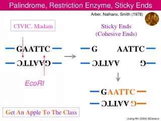
Restriction Enzyme Info. (page 216-218) • rDNA (recombinant DNA)—the produced piece of DNA from inserted another piece of DNA • recognize specific sites to cut the DNA • Blunt ends—straight across • Sticky ends—one side of DNA is longer than the other, these overhangs allow for complementary matches between two DNA pieces cut by the same enzyme, the sticky ends match and pasting ma occur to produce an rDNA molecule • More than 1200 restriction enzymes discovered & isolated from bacteria • Read 5 3 • Palindromic (example radar or GAATTCCTTAAG
Naming of restriction enzymes • Based on their origin and order of discovery • 1st E. coli EcoRI
Why Restriction Enzymes are important:Transforming Cells Uptake and expression of foreign DNA by a cell Making Recombinant DNA
Prep. For RE cleavage lab 1 week before1. label tubes—8 of each DNA EcoRI HindIII 2. Make more TAE buffer Monday • Prepare 0.8% agarose solution (add __g agarose to ___ml of TAE, heat until clear) and then pour gels • Pool DNA—spin in microfuge
Preparing gels • ___ grams agarose • Add up to ___mL buffer • Melt in microwave, let cool • Set up trays—use 6 well comb • Pour about 30-50mL into each tray • Add 1uL ethidium bromide
Each Group will run 3 DNA samples • Vial lambda DNA • Vial lambda DNA cut with EcoRI • Vial Lambda DNA cut with HindIII
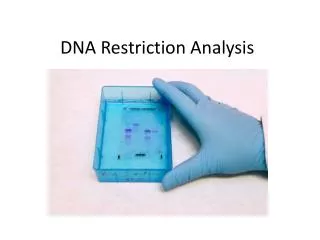
Gel loading—change in protocol Make sure to record what is in each lane in your lab notebook
Results and Discussion • Before you leave the lab make, sure you have your measurements for the table in 4c • Use the graph paper to graph the table in 4c as explained in 4d-4j • Attach the graph paper to your lab write-up