Restriction Enzyme Digestion
Restriction Enzyme Digestion . Zelha Nil Nov-09. Today’s Laboratory Objectives. Results of gDNA experiment: the concentration, purity, and integrity of genomic DNA Digest genomic DNA and plasmids . DNA quantification.

Restriction Enzyme Digestion
E N D
Presentation Transcript
Restriction Enzyme Digestion Zelha Nil Nov-09
Today’s Laboratory Objectives • Results of gDNA experiment: the concentration, purity, and integrity of genomic DNA • Digest genomic DNA and plasmids
DNA quantification • A UV spectophotometer measures the amount of light particular molecules absorb (Proteins at A280; Nucleic Acids at A260) • Lambert-Beer law describes the relationship between absorptivity coefficient and concentration and is given by the following equation: A=εbc Where: b= light path length c=concentration of substance ε=extinction coefficient For DNA the extinction coefficient, ε= 50 ug/ml
DNA quantification • To Quantify your DNA sample: A260 x Dilution Factor x 50 ug/ml= concentration of nucleic acids in a sample using a 1 cm pathlength (DF=200) • To estimate the purity of your sample: A260/A280= ratio of nucleic acids/protein A260/A280= 1.6-1.8 is optimal for DNA
Integrity of genomic DNA • High Quality Genomic DNA >95% DNA will be of high molecular weight, migrating as intact band near the top of the gel Very little evidence of smaller fragments indicated by a smear of many different sized DNA fragments
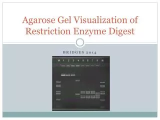
Our results L Z W1 W2 W3 W4 L Z T1 T2 T3 T4 L (1.0% (w/v) agarose, EtBr staining) L: Fermentas GeneRuler™ DNA Ladder Mix; 100-10000 bp DNA ladder Z: Zelha
Restriction Enzymes • Phage (or viruses) invade all types of cells. Bacteria are one favorite target. • Defense mechanisms have been developed by bacteria to defendthemselves from these invasions. • Bacteria have evolved a class of enzymes that destroy foreign DNA (eg. Virus DNA). • protect bacteria from bacteriophages (Viruses). • Infecting DNA is cleaved (restricted) bythe restriction enzyme(s) preventing itfromsuccessfullyreplicatingandparasitizingthecell.
Why the bacteria does not kill itself?The Restriction Enzyme Modification Systems If everything gets cleaved, how come the bacteria does not kill itself? • Usually, organisms that make restriction enzymesalso make a companion modification enzyme(DNA methyltransferase-methylase) that protects their own DNA from cleavage. • These enzymes recognize the same DNAsequence as the restriction enzyme theyaccompany, but instead of cleaving the sequence,they disguise it by methylating one of the bases in each DNA strand.
RE system • This system is composed of a restriction endonuclease enzyme and a methylase enzyme • Each bacterial species and strain has their own combination of restriction and methylating enzymes.
Restriction endonuclease is an enzyme that cuts DNA at internal phosphodiester bonds; different types exist and the most useful ones for molecular biology are those which cleave at a specific DNA sequence.
Classification of restrictionenzymes • Type 1: • Oneenzymewithdifferentsubunitsforrecognition, cleavage, & methylation. • Themethylationandcuttingrxnsbothrequire ATP, Mg+2and S-adenosylmethionine as cofactors. • Theenzymecutsunmodified DNA at somedistance (~1000 bpaway) fromtherecognition site (Asymmetricalrecognitionsequences). • Type 2s: • Asymmetricrecognitionsequence & cleavageoccurs on oneside of recognitionsequenceupto 20 bpaway. • Type 3: • Resembletype 1 systems but havesymmetricalrecognitionsequences.
Type 2: • Restrictionandmodificationaremediatedbyseparateenzymesso it is possibletocleave DNA in theabsence of modification. • Therestrictionactivities do not requirecofactors, makingthemeasiertouse. • Mostimportantly; thoseenzymesrecognize a defined, usuallysymmetricalsequenceandcutwithin it.
Nomenclature • Smith and Nathans (1973) proposed enzyme naming scheme; • Three-letter acronym for each enzyme derived from the source organism • First letter from genus • Next two letters represent species • Additional letter or number represent the strain or serotypes • For example. the enzyme HindII was isolated from Haemophilus influenzae serotype d.
Most type 2 RE recognize and cleave DNA within particular sequences of 4 to 8 nucleotides which have two fold axis of rotational symmetry. Such sequences are often referred as palindromes: • Ex: HaeIII 5’ TGACGGGTTCGAGGCCAG 3’ 3’ ACTGCCCAAGGTCCGGTC 5’
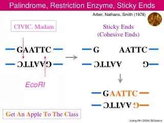
Ends of restriction fragments; • Bluntends • Stickyends • 3‘extensions • 5‘extensions • Importantly, the 5' termini of each strand in thecleavage product(s) retain the phosphoryl group from the phosphodiester bond, the 3' termini are hydroxylated.
AluI HaeIII Blunt ends • Some restriction enzymes cut DNA at opposite base • They leave blunt ended DNA fragments
Sticky ends • Most restriction enzymes make staggered cuts • Staggered cuts produce single stranded “sticky-ends”
Star effect • Optimum conditions are necessary for the expected result. • Under extreme conditions such as elevated pH or low ionic strength, RE are capable of cleaving sequences which are similar but not identical to their recognition sequence. • EcoR1→GAATTC EcoR1 with star activity→NAATTN (N=any base)
General uses of REs • Detection of RFLPs • Restriction enzyme map: The location of the restriction enzyme cleavage sites on the DNA molecule • DNA fragments from different species can be ligated to create Recombinant DNA:
Example Single digest with EcoRI: Double digest with EcoRI & PstI: 6kb 1,5kb 0,9kb 0,8kb 0,6kb 0,2kb 6kb 2,4kb 1kb 0,6kb
Experimental procedure • Genomic DNA isolated last week and the plasmid DNA isolated before will be digested. • Single digestion with EcoRI • Double digestion with EcoRI & HindIII
Group 1 &3: Singledigestion • Group 2&4: Doubledigestion • An Enzymatic Unit (u) is defined as the amount of enzyme required to digest 1 ug of DNA under optimal conditions: 2-3 u/ug of genomic DNA 1 u/ug of plasmid DNA Stocks typically at 10 u/ul