Prothrombin Time (PT)
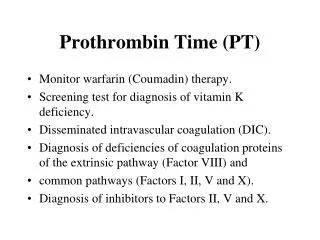
Prothrombin Time (PT). Monitor warfarin (Coumadin) therapy. Screening test for diagnosis of vitamin K deficiency. Disseminated intravascular coagulation (DIC). Diagnosis of deficiencies of coagulation proteins of the extrinsic pathway (Factor VIII) and

Prothrombin Time (PT)
E N D
Presentation Transcript
Prothrombin Time (PT) • Monitor warfarin (Coumadin) therapy. • Screening test for diagnosis of vitamin K deficiency. • Disseminated intravascular coagulation (DIC). • Diagnosis of deficiencies of coagulation proteins of the extrinsic pathway (Factor VIII) and • common pathways (Factors I, II, V and X). • Diagnosis of inhibitors to Factors II, V and X.
Prothrombin Time (PT) • Venous blood is collected in a citrate (blue top) tube. • A mixture of thromboplastin (phospholipid, tissue factor {Factor III} and calcium) added to patient plasma and time to clot formation is determined.
Prothrombin Time (PT) • Common causes of acquired factor deficiencies include severe liver disease and vitamin K deficiency. • Inhibitors to Factor V and X are rare occurring primarily in patients with amyloidosis. • To distinguish factor deficiency from an inhibitor, an inhibitor screen (1:1 mixing study) is performed. If PT corrects, a deficiency should be suspected. If PT does not correct, an inhibitor should be suspected. • Ingestion of warfarin results in non-carboxylated vitamin K-dependent factors resulting in a prolonged PT.
Partial Thromboplastin Time (PTT) • Screening for congenital or acquired deficiencies of intrinsic pathway factors (Factor VIII, VIV, XI, XII, prekallikrein and high molecular weight kininogen. • Monitoring heparin therapy. • Screening for inhibitors of Factor VIII, VIV, and XI. • Screening for lupus anticoagulant. • DIC.
Partial Thromboplastin Time (PTT) • Venous blood is collected in a citrate (blue top) tube. • Mixture of phospholipid (partial thromboplastin) and calcium is incubated with citrated plasma and the time to clot formation is measured. • May be performed as a one-step procedure without an activator or may use an activator such as silica or kaolin (activated PTT or APTT).
Partial Thromboplastin Time (PTT) • Common causes of acquired deficiency of intrinsic pathway factors include severe liver disease and vitamin K deficiency. • Inhibitors to Factor VIII and VIV occur predominantly in severe hemophilia A and hemophilia B respectively. • To distinguish factor deficiency from an inhibitor, an inhibitor screen (1:1 mixing study) is performed. If PTT corrects, deficiency should be suspected. If PTT does not correct, inhibitor should be suspected. • Deficiencies of factors in the common pathway prolong the PT more than the PTT.
1:1 Mixing Study for Prolonged PT • To determine if prolonged PT is due to deficiency of Factor II, V, VII or X or due to the presence of an inhibitor.
1:1 Mixing Study for Prolonged PT • Equal portions of patient plasma and normal plasma are combined and incubated for various lengths of time, typically 0 minutes, 30 minutes, 60 minutes and 120 minutes. • PT is performed. If PT corrects with the addition of normal plasma, a factor deficiency is the cause of the prolonged PT. If the PT corrects partially or not at all, a prolonged PTT is due to the presence of an inhibitor.
1:1 Mixing Study for Prolonged PTT • To determine if a prolonged PTT is due to a deficiency of Factor VIII, VIV, XI, XII, prekallikrein or high molecular weight kininogen or is due to the presence of a factor inhibitor or the lupus inhibitor.
1:1 Mixing Study for Prolonged PTT • Equal portions of patient plasma and normal plasma are mixed and a PTT is performed following incubation at 37 for various times, typically 0 minutes, 30 minutes, 60 minutes and 120 minutes. If the PTT corrects into the normal range, a factor deficiency is indicated. Partial correction or no correction indicates the presence of an inhibitor.
1:1 Mixing Study for Prolonged PTT • Lupus inhibitor is usually evident after immediate mixing, i.e. the PTT is prolonged at 0 minutes and all subsequent time points. • A Factor VIII inhibitor usually corrects or nearly corrects on immediate mixing but fails to correct at later time points (one or two hours).
Fibrin/Fibrinogen Degradation Products (FDP) • Use: • Disseminated intravascular coagulation. • Primary fibrinolysis. • Thrombolytic states. • Collection: • Venous blood is collected in a citrate (blue top) tube or in special clot tubes specifically designated for FDP specimens
Fibrin/Fibrinogen Degradation Products (FDP) • Latex bead agglutination method: latex beads conjugated with antibody recognizing fibrin, fibrinogen, and FDP are incubated with serial dilutions of patient serum. If FDP are present, they will complex with the antibody and cause visible clumping of the latex particles. Automated methods are available. • Serum is prepared by defibrinogenating plasma by the addition of exogenous thrombin.
Fibrin/Fibrinogen Degradation Products (FDP) • FDP may appear to be elevated in patients with congenital or acquired disfibrinogenemias • the reactive material is not FDP but slowly clottable fibrinogen. • Patients with severe liver disease may have elevated FDP due to reduced clearance by the liver. These patients may also have an acquired disfibrinogenemia. Incomplete removal of fibrinogen during the preparation of the sample may yield falsely elevated FDP. Elevated FDP in the absence of DIC may occur in clot lysis, especially in DVT, PE, post-surgical states and portal-caval shunts.
Functional Assays for Factors VIII, VIV, XI, XII, Prekallikrein and High Molecular Weight Kininogen • Use: • Congenital or acquired deficiencies. • Collection: • Venous blood is collected in a citrate (blue top) tube.
Functional Assays for Factors VIII, VIV, XI, XII, Prekallikrein and High Molecular Weight Kininogen • Addition of patient plasma to reagent plasma severely deficient in Factors VIII, VIV, XI, XII, PK or HMWK will correct the prolonged PTT of the factor-deficient plasma. • If the patient plasma contains the factor in question, the level of factor activity in the patient plasma correlates with the extent of correction of the PTT. • The activity is calculated by comparing the correction produced by the patient plasma to the correction produced by various dilutions of normal pooled plasma.
Functional Assays for Factors VIII, VIV, XI, XII, Prekallikrein and High Molecular Weight Kininogen • Decreased Factor VIII is found in hemophilia A, hemophilia A carrier state and some Von Willebrand’s disease. • Increased levels of Factor VIII may occur in inflammatory states, pregnancy and estrogen supplementation. • Decreased levels of Factor VIV found in hemophilia B, hemophilia B carrier state, vitamin K deficiency and severe liver disease. • Decreased level of Factor XI found in hemophilia C and severe liver disease.
Functional Assays for Factors VIII, VIV, XI, XII, Prekallikrein and High Molecular Weight Kininogen • Decreased level of Factor XII found in congenital deficiency of Factor XII, a lupus inhibitor which prolongs the PTT and gives a positive PTT inhibitor screen. • Decreases the level of PTT factors as a group in vitro by its action as a phospholipid inhibitor. • Decreased level of prekallikrein is found in congenital deficiency. • Decreased level of high molecular weight kininogen is found in congenital deficiency.
Functional Assays for Factors II, V, VII and X • Use: • Congenital or acquired deficiencies. • Collection: • Venous blood is collected in a citrate, blue top tube.
Functional Assays for Factors II, V, VII and X • The addition of patient plasma to reagent plasma which is severely deficient in Factors II, V, VII or X will correct a prolonged PT. • If the patient plasma contains the factor in question, the level of factor activity in the test plasma correlates with the degree of correction of the PTT. • The activity is calculated by comparing the correction produced by test plasma to the correction produced by various dilutions of normal pooled plasma.
Functional Assays for Factors II, V, VII and X • Isolated deficiency of Factor II may be congenital or acquired in patients with the lupus anticoagulant. • Isolated deficiency of Factor V may be congenital or due to the effect of an inhibitor in rare cases of amyloidosis (with concomitant Factor X inhibitor). • Isolated deficiency of Factor VII may be congenital or occur in early vitamin K deficiency. • Isolated deficiency of Factor X may be congenital or acquired in patients with primary amyloidosis.
Clot Solubility (Screening Test for Factor XIII Deficiency) • Use: • Screening test for congenital Factor XIII deficiency. • Collection: • Venous blood is collected in a citrate, blue top tube.
Clot Solubility (Screening Test for Factor XIII Deficiency) • Patient plasma is clotted with exogenous thrombin. The clot is incubated with five molar urea for 24 hours or with 1% monochloracetic acid for two hours at 37C. The clot is observed for dissolution. If the patient has less than 2-3% Factor XIII activity, the clot completely dissolves (positive test).
Clot Solubility (Screening Test for Factor XIII Deficiency) • Test is insensitive to mild deficiencies. • A quantitative test (immunologic or biologic) must be used to confirm cases with a positive clot solubility test and to identify mildly deficient patients.
Screening Test for Prekallikrein Deficiency • Use: • Differential diagnosis of a prolonged PTT in an asymptomatic patient includes deficiency of Factor XII high molecular weight kininogen and prekallikrein. • Collection: • Venous blood is collected in a citrate, blue top tube.
Screening Test for Prekallikrein Deficiency • Prekallikrein-deficient plasma has a prolonged PTT. • If the PTT reaction mixture is allowed to incubate for prolonged period (30 mins.) the PTT of a prekallikrein-deficient plasma corrects to normal. This change is not seen in any other factor deficiency.
Fibrinogen Assays • Diagnosis of afibrinogenemia. • Congenital or acquired hypofibrinogenemia or disfibrinogenemia. • Diagnosis and monitoring of DIC. • Monitoring thrombolytic therapy. • Diagnosis of primary fibrinogenolysis
Fibrinogen Assays • Clauss method (dilute thrombin time): Measures the time to clot formation after the addition of thrombin to dilute patient plasma. Fibrinogen value determined from standard curve. • Ellis method: Measures changes in turbidity of undiluted patient plasma following the addition of thrombin. • Clottable protein method: Measures protein mass of clot formed in plasma following the addition of thrombin.
Fibrinogen Assays • Immunologic method: Measures immunologically reactive fibrinogen. • Salt precipitation method: Measures turbidity of plasma following precipitation of fibrinogen by sodium sulfite. • Dupont ACA method: Measures rate of change in turbidity of dilute plasma as fibrin is formed from fibrinogen following the addition of thrombin.
Fibrinogen Assays • Clauss and ACA method may yield falsely low values if specimen contains fibrin degradation products. • Immunologic and Salt Precipitation methods do not reflect functional ability of the fibrinogen and may yield discrepant values when compared with other methods in cases of disfibrinogenemia.
Thrombin Time (TT) • To document presence of heparin. • A prolonged TT which corrects with the addition of protamine sulfate. • A prolonged TT in a sample with a normal reptilase time. • Diagnosis of disfibrinogenemia when performed in combination with a reptilase time. • Monitoring thrombolytic therapy. • Diagnosis of afibrinogenemia or hypofibrinogenemia when performed in conjunction with fibrinogen assays.
Thrombin Time (TT) • Collection: • Venous blood is collected in a citrate, blue top tube. • Principal of test: • Thrombin is incubated with the patient’s plasma and the time to clot is measured.
Thrombin Time (TT) • TT is very sensitive to the presence of therapeutic concentrations of Heparin which may result in a marked prolongation of the TT. • TT is usually prolonged in the presence of increased FDP. • TT is often prolonged in the presence of a paraprotein. • Performance of the reptilase time may be of value in determining the cause of a prolonged TT.
Reptilase Time • Use: • In the presence of a prolonged thrombin time, the reptilase time is useful in diagnosis of dysfibrogenemia, hypofibrinogenemia. • Documenting the presence of heparin. • Collection: • Venous blood is collected in citrate and preserved on ice until the time of testing.
Reptilase Time • Plasma is incubated with reptilase-R and the time to clot formation is measured. • Reptilase-R is a thrombin-like enzyme. • Reptilase-R cleaves only fibrinopeptide A whereas thrombin cleaves fibrinopeptides A & B. • Reptilase R is inhibited only slightly or not at all by heparin and fibrin degradation products.
Reptilase Time • Equally prolonged RT and TT suggest a fibrinogenemia. • Perform fibrinogen determinations functional and immunologic to confirm. • RT is much longer than the TT suggests disfibrinogenemia. • Perform fibrinogen determinations functional and immunologic to confirm. • If the TT is prolonged, and the RT is normal. This suggests heparin is present. • Neutralize heparin with protamine sulfate and repeat TT (TT should normalize). • If TT is prolonged and RT is only slightly prolonged, this suggests FDP. Perform FDP assay to confirm
Quantitative Assay for Factor VIII: C Inhibitor • Use: • Precise quantitation of the amount of Factor VIII C inhibitor in a patient’s plasma. • Collection: • Venous blood is collected in citrate, blue top tube.
Quantitative Assay for Factor VIII: C Inhibitor • Acquired inhibitors to Factor VIII C are quantitated by mixing the patient plasma with a known amount of Factor VIII C (typically the amount present in normal control plasma). • The inhibitor in the patient plasma will bind to and inhibit Factor VIII C activity in a normal control plasma resulting in a decrease in the Factor VIII-C activity. • After an appropriate incubation period, the residual Factor VIII-C activity is measured by comparing the Factor VIII-C activity of the patient/control mixture to that of the control/control mixture.
Quantitative Assay for Factor VIII: C Inhibitor • The amount of inhibitor may be quantitated in Bethesda units. • One Bethesda unit of inhibitor is defined as the amount of inhibitor that will inactivate 50% of the Factor VIII-C activity present.
Ristocetin Co-factor Activity Assay • Use: • Diagnosis of Von Willebrand’s disease. • Collection: • Blood is collected in a plastic syringe containing 3.8% sodium citrate in a ratio of 1:10 anticoagulant to whole blood.
Ristocetin Co-factor Activity Assay • Level of ristocetin co-factor activity is determined by the rate at which patient plasma induces aggregation of a standardized formalin-fixed platelet suspension in the presence of ristocetin (an antibiotic with a high affinity for the receptor for Von Willebrand’s factor). • This test is a functional assay for Von Willebrand’s factor.
Ristocetin Co-factor Activity Assay • Ristocetin co-factor activity is a property predominantly of high molecular weight multimers of Von Willebrand’s factor. • Lack of ristocetin co-factor activity may represent an abnormal distribution of multimers rather than an absolute deficiency of Von Willebrand’s factor.
Ristocetin Co-factor Activity Assay • While ristocetin co-factor is usually deficient in some degree in the plasma of all patients with Von Willebrand’s disease, the ratio of ristocetin co-factor activity to Von Willebrand’s factor antigen level differs among the sub-types. • The level of ristocetin co-factor activity decreases with DDAVP infusion and this assay is one of the methods used to evaluate the therapeutic response to the infusion.
Von Willebrand’s Factor Antigen Assay • Use: • Diagnosis of Von Willebrand’s disease. • Collection: • Blood is collected in a plastic syringe containing 3.8% sodium citrate in a ratio of 1:10 anticoagulant to whole blood.
Von Willebrand’s Factor Antigen Assay • Rocket immunoelectrophoresis and radial immunodiffusion are commonly used techniques. • Rocket immunoelectrophoresis: Patient plasma is placed in a well in agarose gel containing monospecific antibodies against Von Willebrand’s factor antigen. The gel is electrophoresed and the one Von Willebrand’s factor antigen antibody complexes precipitate in a rocket or peak, the height of which is proportional to the antigen content of the sample. • Radial immunodiffusion: Patient plasma sample is allowed to diffuse in an antibody containing gel from a central well. The Von Willebrand’s factor antigen antibody complexes precipitate and the diameter of the ring formed by the precipitate is measured. The diameter of the ring is directly proportional to the amount of Von Willebrand’s factor antigen in the specimen.
Crossed Immunoelectrophoresis for Von Willebrand’s Factor • Use: • Differentiation of types of Von Willebrand’s disease. • Collection: • Blood is collected in a citrate blue top tube.
Crossed Immunoelectrophoresis for Von Willebrand’s Factor • Patient plasma is electrophoresed in a narrow line at the bottom of an agorous gel to first separate the multimers of Von Willebrand’s factor by molecular weight. • Agarose gel containing a monospecific antibody for Von Willebrand’s factor is then applied to a portion of the electrophoresis plate above the line containing the electrophoresed Von Willebrand’s factor and the sample is electrophoresed into the antibody-containing gel in a second dimension 901. Antigen antibody complexes form and precipitate as arcs in the gel. The highest point of the precipitant arc and the shape of the arc indicate the distribution of Von Willebrand factor multimers.
Crossed Immunoelectrophoresis for Von Willebrand’s Factor • Interpretation of the test is qualitative • An increase in high molecular weight multimers may be seen in patients treated with DDAVP.
Tissue Thromboplastin Inhibitor (TTI) • Diagnosis of the lupus inhibitor. • TTI may be positive for the lupus inhibitor even when the PTT is normal. • TTI is often used in conjunction with other tests to detect or confirm the presence of the lupus inhibitor. These include PTT inhibitor screen, anti-cardiolipin antibody test, platelet neutralization procedure, dilute stipend time (dilute Russell’s viper venom time), agorous gel inhibition test and kaolin clotting time.
Tissue Thromboplastin Inhibitor (TTI) • Patient plasma and normal control plasma are incubated with calcium and diluted thromboplastin (1:100 and 1:1,000). • Time to clot formation is measured. • If an inhibitor is present in the patient plasma, the time until clot formation will be significantly greater than that of control plasma. • A ratio of patient to control clotting time is calculated. • A ratio of greater than 1.2 is considered positive for an inhibitor.