Download
1 / 56
630 likes | 1.15k Views
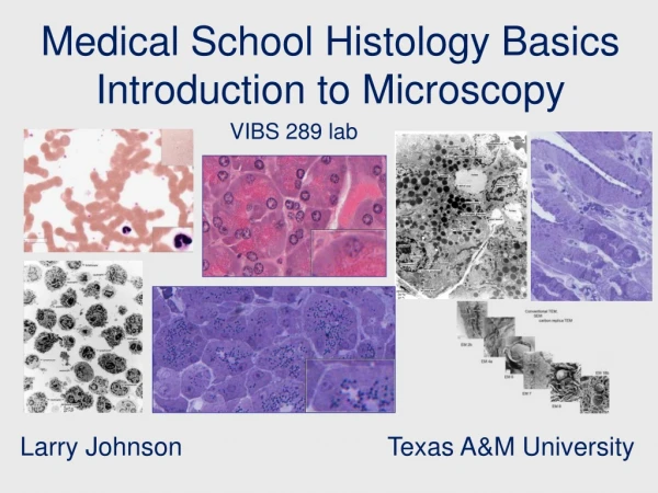
Introduction to Histology. Al- Maarefa College. Objectives. Understand what is Histology Realize the size of histological tissues Know basic steps in tissue preparation and staining Understand the importance of different stains Identify different techniques of imaging in histology.

E N D
Introduction to Histology Al-Maarefa College
Objectives • Understand what is Histology • Realize the size of histological tissues • Know basic steps in tissue preparation and staining • Understand the importance of different stains • Identify different techniques of imaging in histology
What is Histology? • The name "Histology" is derived from Greek: • "Histos“: tissue • "logos" : the study of • Histology: the study of tissues
What is Histology? • The study of the microscopic anatomy of cells and tissues • How? • Examining a thin slice (section) of tissue under a light microscope or electron microscope • Enhanced by using different histological stains
Levels of body organization • Cells: • The smallest independently living highly complex structure • Tissues: • Functional unit: group of cells of similar function and origin (with matrix between cells) • Organs: • Several tissues grouped together • Systems: • Several organs working for a common function
Tissues • Tissues are made of: • cells and • extracellular matrix • Intense interaction between cells and matrix • Cells and extracellular matrix form a continuum that functions together and reacts to stimuli and inhibitors together
Tissues • Four fundamental tissues are recognized: • Epithelial tissue • Connective tissue • Muscular tissue • Nervous tissue
The size spectrum • The small size of cells and matrix components makes histology dependent on the use of microscopes
Light MicroscopyWith a maximum magnification power of x 1000 times
Electron MicroscopyWith a maximum magnification power of x 1000000 times
Electron Microscopy Surface of epithelial cell, X 100,000 Bronchiole, epithelial cells, X 5000 www.visualhistology.com
Basic Techniques • Preparation of histological sections • Fixation • Processing (dehydration and clearing) • Embedding • Cutting • Staining • Permanent Mounting
Preparation of histological sections • Fixation • Fixing tissues by chemicals so they will not change their volume and shape during processing • Keeps tissue as close to their living state as possible • prevents autolysis and bacterial attack • prepares tissues for staining • Fixatives: • Acetic acid, Formaldehyde, Ethanol, Glutaraldehyde, Methanol and Picric acid.
Preparation of histological sections • Dehydration and clearing • removes fixative and water from the tissue and replace them with dehydrating fluid • Uses hydrophilic or alcohol solutions to extract water • Examples of solutions: • Ethanol, Methanol, Acetone
Preparation of histological sections • Embedding • is the process by which tissues are embedded in a medium (agar, gelatin, or wax) which when solidified provides sufficient external support during sectioning
Preparation of histological sections • Cutting • Small (micro) sections are cut by a Microtome • Section thickness • 2 to 25 micrometers=um thick for light microscope • 60 to 100 nanometers thick for electron microscopy
Preparation of histological sections • Staining • Various stains are used to see and highlight specific structures and molecules
Preparation of histological sections • Staining • Various stains are used to see and highlight specific structures and molecules
Hematoxylinand Eosin (H & E) • H & E stains are universally used for routine histological examination of tissue sections.
Staining Techniques • Hematoxylin and Eosin (H&E) • Hematoxylin is a basic dye that stains acidic components of cells a blue (basophilia) • Stains the nuclei of cells, and the rER of cytoplasm
Staining Techniques • Hematoxylin and Eosin (H&E) • Eosin is an acidic dye that stains the basic components of cells reddish-pink (acidophilia) • Most of cytoplasm of cells is stained by eosin
Hematoxylinand Eosin (H & E) • Nuclei - blue - with some metachromasia • Cytoplasm - various shades of pink-identifying different tissue components
Hematoxylin Blue colored Positively charged (colors negatively charged molecules, such as DNA, rER) The ability of such anionic groups to react with a basic dye - basophilia Eosin Red colored Negatively charged (colors positively charged molecules (amino acids, cytoplasm) The ability of such cationic groups to react with an acidic dye - acidophilia Features of H&E
Staining Techniques • Periodic acid-Schiff (PAS) staining • mucus, basal lamina, glycogen (carbohydrates) H&E stain does not preserve Mucous in Goblet cells, which Appear empty PAS stain preserves Mucous in Goblet cells, which and stains it with a magenta colour Wheater’s Functional Histology, a text and colour atlas Junqueira and Carneiro, Basic Histology, a text and atlas
Staining Techniques • Periodic acid-Schiff (PAS) staining • mucus, basal lamina, glycogen (carbohydrates) Glycogen
Staining Techniques • Periodic acid-Schiff (PAS) staining • mucus, basal lamina, glycogen (carbohydrates) Carbohydrate content in liver tissue stained magenta with PAS stain Clarke F. Millette, Univ. of South Carolina, USA
Staining Techniques • Toluidine blue (Metachromatic stain) • Blue stain that stains specific components of tissues a purple color • Metachromasia is seen in the matrix of hyaline cartilage, or in the granules of mast cell IJDVL, Omar El Safoury Science Photo Library Blue stain of Hyaline cartilage Metachromasia of Mast cells, skin
Staining Techniques • Oil Red O Stain lipids red-orange in unfixed frozen sections • Sudan black Stains lipids black in unfixed frozen sections
Staining Techniques • Impregnation Silver impregnation techniques are also widely used to demonstrate reticular fibers Lymph node, Silver Impregnation Seoul National University
Staining Techniques • Reticulin Reticular fibers are stained dark. Used esp. in liver Smooth Muscle, Reticulin stain Clarke F. Millette, Univ. of South Carolina, USA
A liver biopsy stained using the reticulin demonstrating the normal hepatic plate thickness.
Staining Techniques • Giemsa stain • Blood smears, for disease PlasmodiunFalciparum (Malaria) Trypanosoma sp. CDC /Dr. Myron G. Schultz 1970 www.sciencebuzz.org
Preparation of histological sections • Mounting • preserves and supports a stained section • mounted on a clear glass slide, and covered with a thin glass cover-slip
Basic Techniques • Total preparation time: 16 hours • Immediate Frozen section time: 5 minutes • For immediate results – (in tumor surgery) • Results not as accurate as formalin fixed, wax embedded specimens
Basic Techniques • In some cases the tissue to be examined is a very thin membrane (Cell Smears) • blood or bone marrow http://hepatitiscresearchandnewsupdates.blogspot.com
Basic Techniques • In some cases the tissue to be examined is a very thin membrane (Cell Smears) • blood or bone marrow • epithelial cells • (e.g. from the oral cavity, cervix uteri) Crvical carcinoma, PAP stain Univ of Uklahoma Health Science Center NATIONAL CANCER INSTITUTE / SCIENCE PHOTO LIBRARY
Some definitions • Histology: • microscopic study of tissues • Biopsy: • removal of tissues for diagnostic purposes • Autopsy: • examination of organs of a dead body (to find clues to determine cause of death)
Phase-Contrast Microscopy • An optical microscopy illumination technique • A small phase shifts in the light passing through a transparent specimen are converted into amplitude or contrast changes in the image. • Does not require staining to view the slide Epithelial cell from cheek
Polarizing Light Microscopy • Polarization is a property of Electromagnetic waves, such as light that describes the orientation of their oscillations Polarizer is rotated to transmit the reflections as well as possible • By rotating the polarizer by 90° , almost all reflected sunlight is blocked
Polarizing Light Microscopy • Polarization is a property of Electromagnetic waves, such as light that describes the orientation of their oscillations
Polarizing Light Microscopy • Collagen fibers • Yellow birefringence
Fluorescence Microscopy • A sample is illuminated with light of a one wavelength which causes fluorescence in the sample. The light emitted by fluorescence, which is at a different, longer, wavelength than the illumination, is then detected through a microscope objective
Fluorescence Microscopy • Epifluorescent imaging of the three components in a dividing human cancer cell. • DNA is stained blue, • a protein called INCENP (Inner centromere protein) is green, and • the microtubules are red. • Each fluorophore is imaged separately using different filters, and images are captured sequentially using a digital CCD camera( charge coupled device), then overlaid to give a complete image.
Scanning Electron Microscopy Clarke F. Millette, Univ. of South Carolina, USA
Scanning Electron Microscopy Bruce Wetzel/Harry Schaefer, courtesy National Cancer Institute