Download
1 / 1
10 likes | 196 Views
Chi-Sheng Chien 1 , Tze-Yuan Liao 2 , Tsung-Yuan Kuo 2 , Ming-Yang Kuo 1 , Tzer-Min Lee 3.

E N D
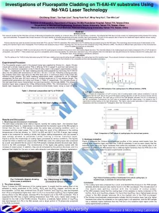
Chi-Sheng Chien1, Tze-Yuan Liao2, Tsung-Yuan Kuo2, Ming-Yang Kuo1, Tzer-Min Lee3 1Division of Orthopedics, Department of Surgery, Chi-Mei Foundation Hospital, Tainan 710, Taiwan,China2Department of Mechanical Engineering, Southern Taiwan University, Tainan 710, Taiwan, China3Medical College Institute of Oral Medicine, National Cheng Kung University, Tainan 710, Taiwan, China AbstractINTRODUCTIONThis research studies the Nd-YAG laser process of fabricating fluorapatite (FA) cladding on a titanium alloy, which are used widely in medicine. The adopted Nd-YAG laser process results in a metallurgical bonding between the coating layer and substrates. Fluorapatite was chosen because of its higher stability at elevated temperatures and lower solubility than hydroxyapatite. The aim of this research was to improve the traditional implant material, whose coating layer can dissolve or deteriorate in a human body after long periods of time. METHODSPolyvinyl alcohol was used as a binder and was mixed with FA to coat the Ti-6Al-4V substrates. After being dried, the FA cladding was processed using the Nd-YAG laser. The morphology and microstructure of weld beads on the coating and transition layers were investigated. Their hardness was analyzed using a Vickers hardness test. The structures were analyzed using X-Ray Diffraction (XRD). The effects of different laser parameters on the characteristics of the FA cladding properties are discussed.RESULTSAt a laser power of 740W and 1150W, the microstructure of the FA coating layers consisted of the porous coral- and needle-shaped structure. The chemical phases in the coating layers were mainly composed of FA, β-TCP (tricalcium phosphate), and Ca2P2O7. These phases have good bio-compatibility and bio-decomposability. No cracks were observed in any of the samples when the indentation test was performed at the interface between the coating layer and the transition layer, which indicates a satisfactory adhesion.CONCLUSIONThe FA coating on the Ti-6Al-4V alloy fabricated using Nd-YAG laser cladding had a good bonding strength between the coating layer and the transition layer. The produced chemical compound possessed porous structures and allowed for the formation of bio-compatible and bio-decomposable bone tissue. Investigations of Fluorapatite Cladding on Ti-6Al-4V substrates Using Nd-YAG Laser Technology Fig.3 XRD analysis of the coating layers for different binders .(740W) 3. Ca/P ratio of coating layers Figure 4 reveals the comparison of Ca/P (in wt.%) ratio of coating layers under various conditions. It can be seen clearly that the Ca/P ratio of the current FA-based coatings depends on both the choice of binder and the laser processing parameters. The Ca/P ratio is 3.6-5.8 in the PVA samples, whereas 13.6-15.1 is observed in the WG counterparts. Furthermore, the Ca/P ratio is increased with the output power. It is related to the P vaporization during heating at the high temperature. The higher output power generates a higher processing temperature and thus increases the P dissipation effect from the structure. Table 2. Parameters used in Nd-YAG laser cladding process. Results and Discussion 1. Morphology and microstructure of weld beads The weld bead comprises three distinct regions, namely the coating layer , the transition layer and substrate (Figure 1)., respectively. It is observed that the transition layer of the WG sample is thinner than the one of PVA sample under the same laser processing condition, and it is increased with the output power. This is most likely the result of the difference in the melting temperatures of the two binders, i.e. 1300C for WG and 240C for PVA. A larger input energy needs to be consumed for melting a binder with higher melting temperature, and this causes a less energy allowed to penetrate further forming a thinner transition layer. Figure 2 shows the morphology of coating layers processed at various conditions. The figures reveal that when a relative low output power (740W) was used, the coating layer was composed by a coral- and dendrite-like structure in the PVA sample; whereas dendrite structure was mainly found in the WG counterpart. The microstructure of the layers was getting coarser with the increase of output power. In addition, they are all composed by fine porous structures, and it is believed that helpful for Ca ions or bone cells attraction and attachment following implantation. Experimental Procedure The Fluorapatite powers used in the present study was supplied by Showa Inc., Japan. FA was mixed with a binder material (either PVA or WG) by 50:50 in weight % and then well stirred into slurry. The chemical composition of the Ti-6Al-4V alloy used in the present experiments is shown in Table 1. Substrates for the laser cladding process were prepared by machining the Ti-6Al-4V alloy into thin plates with dimensions of 100 mm 60 mm 3.8 mm. Following a drying process, the samples were laser clad using an Nd-YAG laser set to a continuous wave mode under two different output powers. The laser cladding experiments were conducted in an Ar shielded atmosphere (Ar flow rate: 25 l/min) using a 5 incident angle and a 15 mm defocus length. The experimental parameters and setup are shown in Table 2, respectively. The microstructures of the clad specimens were characterized via scanning electron microscopy (SEM). The phases of transition layer were analyzed by X-Ray diffractometry (XRD). The microhardness of the weld beads was measured by a Vickers automaticmicrohardness tester using a load of 300g. Table 1. Chemical composition (wt-%) of Ti-6Al-4V Fig.4 Comparison of Ca/P values of coating layers for various laser powers. 4. Hardness evaluation Figure 5 presents the Vickers microhardness profiles of the various weld beads from the coating layer, transition layer and then into Ti-6Al-4V substrate. It can be seen clearly that the transition layer has a far higher hardness than either the substrate or the coating layer in all specimen. In addition, the WG sample has a relatively higher hardness than the PVA one in the transition layer region under the same experimental conditions. Fig.5 Vickers hardness profiles of weld beads from central coating layer, to transition layer, to substrate for various laser powers. Fig.1 Schematic diagram showing three zones in weldment. Fig .2 Morphology of coating layers processed at various conditions. • Conclusion • The coating layer was composed by a coral- and dendrite-like structure in the PVA sample; whereas dendrite structure was mainly found in the WG counterpart. The microstructure of • the layers was getting coarser with the increase of output power. • 2.The XRD analysis reveal that the coating layer of the samples is mainly composed of FA, CaTiO2, Al2O3 and Ca3(PO4)2 phases. It is found that Ca3(PO4)2 has a highest peak among others in the WG sample, whereas FA is the highest one in the PVA counterpart. • 3.The Vickers microhardness measurements indicate that the transition layer has a far higher hardness than either the substrate or the coating layer in all specimen, and the WG sample has a relatively higher hardness than the PVA one in the transition layer region under the • same experimental conditions. 2. The phase analysis Figure 3 shows the XRD spectrum of the coating layers. It reveals that the coating layer of the samples is mainly composed of FA, CaTiO2, Al2O3 and Ca3(PO4)2 phases, and they are all biocompatible. CaTiO2 was combined from FA and Ti-6Al-4V, and Ca3(PO4)2 was decomposed from FA at a temperature higher than 1057C [17]. It is found that Ca3(PO4)2 has a highest peak among others in the WG sample, whereas FA is the highest one in the PVA counterpart. This is supposed that WG has a higher melting temperature than PVA, and a higher laser energy is required for the melt of the FA-WG mixture. Therefore, a relatively higher portion of Ca3(PO4)2 was decomposed from FA in the WG sample, and a larger portion of FA phase was preserved in the PVA counterpart. Southern Taiwan University Materials & Micro-Joining Laboratory