Download
1 / 46
470 likes | 679 Views
Recognizing Different Sports Injuries and The Healing Process. Key Terminology. Key Terminology. Swelling - Enlargement of organs, skin, or other body parts; Caused by the build up of fluid in the tissues Ecchymosis – black/blue discoloration due to hemorrhaging

E N D
Recognizing Different Sports Injuries and The Healing Process
Key Terminology • Swelling- Enlargement of organs, skin, or other body parts; Caused by the build up of fluid in the tissues • Ecchymosis – black/blue discoloration due to hemorrhaging • Edema – the collection of fluid in connective tissue • Inflammation- A basic way in which the body reacts to infection, irritation or other injury
Key Terminology • Modality - A therapeutic method or agent • Analgesic – agent that relieves pain w/out causing a complete loss of sensation • Vasoconstriction – decrease in the diameter of the blood vessel • Vasodilation – Increase in the diameter of the blood vessel • Erythema – redness of the skin
Pain and Acute Injury • Everyone copes with pain differently. • Pain is as much psychological as physiological. • Pain results from sensory input received through the nervous system and indicates location of tissue damage. • Pain is not a useful indicator of injury severity.
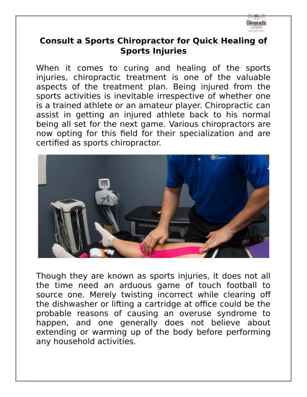
Modalities • Cryotherapy • Appropriate immediately following an injury (20min) • Direct application of cold causes vasoconstriction in the affected tissues • Constriction of the surrounding blood vessels • Why?? • Analgesic – decreases pain • reduces muscle spasm (involuntary contraction of muscle) • Decreases blood flow, decreases temperature = a decrease in secondary swelling • Ice bags or packs, aerosol coolants, ice cups, ice water immersion/whirlpool
Modalities • Thermotherapy • Appropriate 48-72 hours post injury • Considered an analgesic • Why should you not use heat for an acute injury? • Increases blood flow, Increases temperature = anIncrease in swelling = longer recovery time • Your goal is to decrease swelling when dealing with an acute injury • Hydrocollator packs, paraffin wax, warm whirlpool and ultrasound
NSAIDs • Non-Steroidal Anti-Inflammatory Drug • NSAIDs are very popular drugs. • They work to decrease inflammation • Common NSAIDs include Aspirin, Ibuprofen, Motrin, Aleve, and Advil • Rx (prescription) or OTC (over the counter)
Acute or chronic in nature • Acute • Result of sudden trauma; Isolated event • Examples??? • Chronic • Caused by repetitive, overuse activities • Examples???
Acute Traumatic Injuries Fractures • Result of extreme stress and strain on bone – breaks bone • Bone Anatomy • Diaphysis -shaft • hollow and cylindrical • Epiphysis - composed of cancellous bone and has hyaline cartilage covering • Periosteum - dense, white fibrous covering • contains blood vessels and osteoblasts
Acute Fractures • Partial or complete • Either closed or open (through skin) • Presents with deformity, point tenderness, swelling and pain with movement
Load Characteristics • Bones can be stressed or loaded through: • Tension • Compression • Bending • twisting • shearing • Greater force = more severe fracture • Some bones will require more force than others
Mechanical Forces of Injury • Tendons resist tensile forces. • Bones resist compressive forces. • Ligaments resist tensile forces. Each type of tissue has a limit for how much force it can withstand.
Healing of a Fracture • Generally requires immobilization (boot or cast) • usually 6-8 wks • Following cast removal, normal stresses and strains will aid in healing = Rehabilitation
Stress Fractures (fx) • Possible causes • Overload due to muscle contraction, • Altered stress distribution due to muscle fatigue • Changes in surface • Too much activity too soon or in general • Begins with a dull ache becoming worse over time • Initially pain during activity and then progresses to pain following activity • Earlydetection is difficult, bone scan is useful • X-ray is effective after several weeks • If suspected, Stop activity and refer to Orthopedic
Dislocation’s and Subluxation’s • Dislocations • At least one bone in a joint is forced completely out of the respected joint • Most common at the fingers, ankle, and shoulders • Subluxations • Partial dislocation of the joint. Usually reducing itself because the bone did not come completely out of the joint. • Often occurs at the shoulder and patella • Signs and symptoms • Deformity • Swelling • Subluxation – pain and the sensation that it ‘came out’ of place
Signs and symptoms continued… • Other factors associated with dislocations – 1) loss of limb function 2) swelling and point tenderness • Additional concerns • Avulsion fractures, ligament/muscular damage • “Once a dislocation, always a dislocation” • Treatment (tx) • Reduction- generally done by a Physician • Dislocations (particularly first time) should always be considered and treated as a fracture until ruled out • X-ray is a MUST • Return to play often determined by extent of soft tissue damage
Ligament Sprains • Damage to a ligament (provides support to a joint) • Connects a bone to another bone • Extreme twist or rotation of the joint results in the stretching or tearing of the ligament
Grading System • Grade/degree 1 – Stretching of the Ligament • some pain, minimal loss of function, no abnormal motion, and mild point tenderness, slight swelling and joint stiffness • Grade/degree II – Stretching and some tearing of ligament • pain, moderate loss of function, swelling, and instability, some tearing of ligament fibers • Grade/degree III – Tearing/rupture of the ligament • Severe pain, significant loss of function, severe instability and swelling, and may also represent subluxation
Ligament Sprains • Restoration of joint stability is difficult with grade I and II injuries • Must rely on other structures around the joint • Rely heavily on muscles surrounding joint • Ligament has been stretched/partially torn causing development of inelastic scar • Ligament will not regain original tension Rehab = strengthening = improved joint stability
Contusions (Bruise) • Result of sudden blow to body, blunt force trauma • Deep or superficial • Hematoma = blood and lymph flow of tissue • bleeding results in discoloration of skin • Must be cautious of repeated blows to same area • Myositis Ossificans • Calcium deposits form within the soft tissue • Protect the area with padding • Quadriceps and biceps are common sites
Muscle Strains • MOI • Stretch, tear or rupture of muscle or connective tissue • Signs and Symptoms • Hear/feel a pop - Tightness • Deformity - feels like they have been • Pain ‘shot’ • Decreased ROM/flexibility • Rehabilitation • Lengthy process regardless of severity • 6-8 weeks • Return to activity too soon= re-injury
Grades/severity • Grade/Deg I - some fibers have been stretched or actually torn resulting in tenderness and pain on active ROM; movement painful but full ROM present • Grade/Deg II – more fibers have been torn and contraction is painful; usually a depression or divot is palpable; some swelling and discoloration are present • Grade/Deg III- Complete rupture of muscle or musculotendinous junction; usual loss of function; initial great deal of pain that diminishes due to nerve damage
Muscle Cramps • Painful involuntary contraction • Attributed to dehydration/electrolyte imbalance and fatigue Muscle Soreness • Overexertion in strenuous exercise resulting in pain • Two types of soreness • Acute-onset muscle soreness - accompanies fatigue, and is transient muscle pain experienced immediately after exercise • Delayed-onset muscle soreness (DOMS) - pain that occurs 24-48 hours following activity that gradually subsides (pain free 3-4 days later) • Caused by slight microtrauma to muscle
Nerve Injuries • MOI • Two main causes • compression and tension • Acute or Chronic • Causes pain and result in sensory responses • pinch, burn, tingle, muscle weakness, radiating pain • Minor → Severe → Life Altering • Healing process is very slow and long term • Tx – • Referral, rest, Anti-inflammatories • No sport related activity until asymptomatic
Chronic Overuse Injuries • Constant Inflammation • Essential part of healing process • Must occur following tissue damage to initiate healing • Signs and Symptoms • Pain, • Redness • Swelling • Loss of function • Increase in Temperature • If source of irritation is not removed then inflammatory process becomes chronic
Tendinitis • Most common overuse injury • S/S • swelling and pain • Decreased function and ROM • May also experience crepitus • Key treatment = rest • Maintain fitness through non-weight bearing (NWB) or non impact activities • Swimming, bike and elipitical
Tenosynovitis • Inflammation of synovial sheath • Area of the tendon that’s subject to ↑ of friction • Acute = sudden onset, crepitus, and diffuse swelling • Chronic = thickening of tendon with pain and crepitus • Common in the long flexor tendons of fingers • Tx • same as Tendinitis • NSAID’s
Bursitis • Bursa • Fluid filled sac found in areas of friction • Knees, shoulders, ankles, elbows, etc • Acute bursitis • Sudden traumatic injury • Chronic bursitis • Overuse and constant external trauma • S/S • Swelling = increase in pressure due to limited space around anatomical structures • Pain, and some loss of function
Importance of the Healing Process Following Injury • Must understand the sequence and time frame of the various phases of healing • Interference with healing process will delay return to full activity • Need optimal healing environment • Little can be done to speed the process, while much can be done to impede it
3 Phases • Inflammatory • Fibroblastic (Repair) • Maturation - Remodeling
Inflammatory Phase: 5 Cardinal signs of Inflammation • Swelling • Loss of function • Pain • Reddening of skin (erythema). • Warmth – an increase in temperature of the affected area.
Factors that interfere with Healing • Severity of injury • Swelling • Muscle spasm • Atrophy • Infection • Age • Health/nutrition
Inflammatory Phase • Begins immediately following injury • Without the inflammatory phase the other phases will not occur • Vasoconstriction initially, followed by vasodilation • Damage to blood vessels results in blood flow into interstitial spaces causing a hematoma. • Blood clot if formed to stop bleeding • Plasma proteins, platelets, and leukocytes move out of capillaries and into damaged tissue. • Leukocytes engage in phagocytosis • Phagocytosis occurs to clean the injured area • ‘eats’ the debris and injured tissue
Inflammatory Phase • Chemical mediators are released to facilitate healing • Symptomatically presents with the 5 cardinal signs of inflammation • The acute inflammatory process results in a walling off of the damaged area from the rest of the body. • The process acts to clean up the debris and provide components for healing. • Inflammation phase lasts 2-4 days
Fibroblastic Repair Phase • Proliferative and regenerative activity occurs resulting in scar formation • Special leukocytes migrate to the area which break down debris and prepare the area for regeneration and repair. • Unorganized development of scar tissue/collagen is laid down • The collagen fibers are laid down randomly • As the tissue continues to proliferate the area of injury becomes stronger • Therefore the tensile strength of wound ↑rapidly in proportion to collagen synthesis • This signals the beginning of the next phase
Fibroblastic Repair Phase • Occurs within initial hours of injury and continues up to 4-6 weeks • S&S of inflammatory phase subside • Athlete will still experience some tenderness and pain with motion • With development of scar - complaints of pain and tenderness will decrease • Scar tissue can be 95% as strong as the original tissue. Stress on the tissue is helpful for rehabilitation; exercises are critical to this process.
Maturation-Remodeling Phase • Long-term process • Re-alignment of scar tissue according to tensile forces acting on tissue • Re-align to position of maximum efficiency (parallel to lines of tension) • Tissue gradually resumes normal appearance and function • After 3 weeks • Firm, strong, contracted, nonvascular scar exists • Maturation may take several years to be totally complete