Download
1 / 38
380 likes | 536 Views
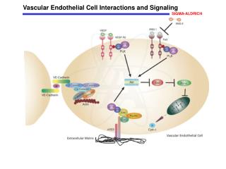
Vascular prothesis material modification to enhance endothelial cell adhesion. T. Markkula, F. Pu, R.L. Williams, J.A. Hunt. Department of Clinical Engineering University of Liverpool. L929 fibroblasts on PET.

E N D
Vascular prothesis material modification to enhance endothelial cell adhesion T. Markkula, F. Pu, R.L. Williams, J.A. Hunt Department of Clinical Engineering University of Liverpool
L929 fibroblasts on PET PET surface cleaned ultrasonically for 30 min with 70 % Ethanol and for 30 min with water prior to cell culturing
Vascular grafts Replacement of blood vessels with artificial implants PTFE and PET most commonly used >6 mm Ø prosthesis OK Smaller grafts thrombosis
Improvements • Find a new material • Modify existing materials • Engineer new tissue • Endothelial cell lining of inner surface of prosthesis
What we did: • Plasma treatment of polymer surface • Endothelial cells seeded on the new surface
Endothelial cells • Seeded on the surface in vitro before operation • Importance of adhesion to graft material
Problems • In vivo endothelial cells become detached inflammation thrombosis • Role of leucocytes in detachment process?
Endothelial cell adhesion Macrophages Endothelial cells Material surface
Improvements • Modify the surface to become more ‘endothelium friendly’ • Improved adhesion is not enough. Cells need to stay on surface even in vivo. • Try to change interaction of endothelial cells with inflammatory cells through surface modification
Endothelial cell adhesion Macrophages Endothelial cells Material surface
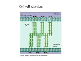
Immuno-globulin superfamily • Integrins • Selectins Adhesion molecules
Endothelial cells to other cells 3 • Endothelial cells to Endo-thelial cells 2 • Endothelial cells to material surface 1 Interactions
Materials • PET poly(ethyleneterephthalate) -[CH2-CH2-O-C- -C-O]n- O O • PTFE poly(tetrafluoroethylene) -[CF2-CF2]n-
RF-plasma systems Gases • Ammonia - NH3 • Nitrogen - N2 • Oxygen – O2 • Argon - Ar • Nitrous oxide - N2O • Air • Inductive coil glass tube 3 W • Capacitor plate glass barrel 80 W Treatment times • 1 - 30 min
Surface analysis • Surface chemistry - XPS, SIMS • Wettability - DCA • Surface morphology - AFM
Surface characterization results • Range of wettabilities and chemistries • Wettability does not necessarily follow the introduction rate of O and N on the surface
Interactions 3 2 1
Material surface properties Interaction with endothelial cells Endothelial cells (EC) interacting EC interacting with blood cells Thrombosis or no
Cell culture analysis • Cellular interaction by expression of adhesion molecules (Flow cytometry, FACS) (immunohistochemistry) • Cell numbers and morphology
Endothelial cell adhesion In vitro cell culturing Plasma treated PTFE Untreated PTFE Plasma treated PET Untreated PET PS cover slip (control)
Endothelial cell adhesion • Cell culturing of endothelial cells alone • Co-culture of endothelial cells with macrophages • Endothelial cells express adhesion molecules depending on external stimuli
Flow cytometry (FACS) Mouse antihuman monoclonal antibodies conjugated with FITC, RPE and CyC were used to target CD31, CD54, CD51/61, CD106, CD62E, CD62P and CD62L.The isotope IgG1-k was used for negative control Immunohistochemistry ABC immunostaining protocol was used to visualise the quantified expression.
Expression of adhesion molecules of endothelial cells on PET and PTFE
Immunohistochemical staining Untreated PET NH3-plasma treated PET CD54 - ICAM, P1, D1
Immunohistochemical staining NH3-plasma treated PTFE Untreated PTFE CD54 - ICAM, P1, D1
Cell growth conclusions • Plasma treatment of PET and PTFE with ammonia appeares to be a powerful method to enhance cell attachment • The modification of PET and PTFE slightly alter the profile of adhesion molecules expressed but not significantly
What will be done... • Surface chemistry of samples will be determined using CHEMICAL DERIVATIZATION with XPS... • The whole range of treatments will be tested with endothelial cell / macrophage co-cultures
What wasn’t presented here... • Plasma treatment alters the attachment of macrophages to endothelial cells... • Macrophage numbers, attachment site and endothelial cell adhesion molecule expressions are altered