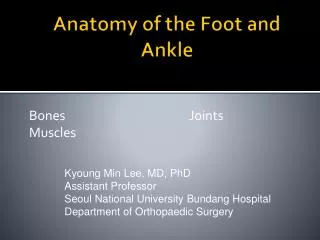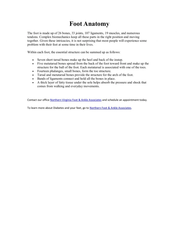
Foot Anatomy
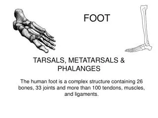
Foot Anatomy. Bone Anatomy. Tarsal Bones Calcaneus Cuboid Navicular 3 Cuneiforms 5 metatarsals 14 phalanges (proximal, middle, distal) Exception. Mnemonic for Learning Tarsal Bones:. T iger C ubs N eed M I L C. N avicular A boat It sails on the Cs. T alus. M edial

Foot Anatomy
E N D
Presentation Transcript
Bone Anatomy • Tarsal Bones • Calcaneus • Cuboid • Navicular • 3 Cuneiforms • 5 metatarsals • 14 phalanges (proximal, middle, distal) • Exception
Mnemonic for Learning Tarsal Bones: Tiger Cubs Need M I L C Navicular A boat It sails on the Cs Talus Medial cuneiform (1) Intermediate cuneiform (2) Lateral cuneiform (3) Calcaneus Cuboid Click R Button for Slideshow
Division of the Foot • Rearfoot • Midfoot • Forefoot
Hindfoot (Rearfoot) • Subtalor Joint • Talus and calcaneus articulation • Calcaneus • Inferior Talus
Midfoot • Composed of • Navicular • 3 cuneiforms • cuboid
Forefoot • 5 MT’s • Proximally 1-3 articulate with cuneiforms • Proximally 4-5 articulate with cuboid • Bases articulate with: • Phalanges
Articulations and Ligamentous Support • Subtalor Joint • Three facets • Motions of the Subtalor Joint • Inversion • Eversion
Hindfoot Articulations and Ligamentous Support • Subtalor Joint • Ligamentous Support Medial Deltoid Ligament Lateral ATF CF PTF • Intra-articular Ligaments • Interosseous Talocalcaneal • Medial Talocalcaneal • Lateral Talocalcaneal
Midfoot Articulations and Ligamentous Support • Six Joints • Talocalcaneonavicular • Calcaneocuboid • Cuboideonavicular • Intercuneiform • Cuneocuboid • Cuneonavicular
Midfoot Articulations and Ligamentous Support • Ligamentous Support • Talocalcaneonavicular Joint • Plantar Calcaneonavicular (Spring Ligament) • Talonavicular • Bifurcate • Calcaneonavicular • Calcaneocuboid • Calcaneocuboid Joint • Bifurcate Ligament • Calcaneocuboid portion • Plantar Calcaneocuboid • Long Plantar Ligament
Midfoot Articulations and Ligamentous Support • Ligamentous Support • Talocalcaneonacicular Joint • Plantar Calcaneonavicular (Spring Ligament) • Talonavicular • Bifurcate • Calcaneonavicular • Calcaneocuboid • Calcaneocuboid Joint • Bifurcate Ligament • Calcaneocuboid portion • Plantar Calcaneocuboid • Long Plantar Ligament
Midfoot Articulations and Ligamentous Support • Ligamentous Support • Intercuneiform Joints • Dorsal and Plantar Intercuneifrom Ligaments • Cuneocuboid • Plantar and Dorsal Cuneocuboid Ligaments • Cuneonavicular Joints • Plantar and Dorsal Cuneonavicular Ligaments
Midfoot Articulations and Ligamentous Support • Ligamentous Support • Intercuneiform Joints • Dorsal and Plantar Intercuneifrom Ligaments • Cuneocuboid • Plantar and Dorsal Cuneocuboid Ligaments • Cuneonavicular Joints • Plantar and Dorsal Cuneonavicular Ligaments
Forefoot Articulations and Ligamentous Support • Tarsometatarsal Joint (Lisfranc’s Joint) • Intermetatarsal Joint • Metatarsalphalangeal Joint (MTP) • Interphalangeal Joint • PIP • DIP
Forefoot Articulations and Ligamentous Support • Ligamentous Support • Intermetatarsal Joint • Plantar Metatarsal Lig • Dorsal Metatarsal Lig • MTP Joints • Plantar Fascia • Plantar Ligament • MCL and LCL • Interphalangeal Joints • Plantar and dorsal joint capsule • MCL and LCL
Arches • Ligaments in foot & ankle maintain arches • Two longitudinal arches • Medial longitudinal arch - extends from calcaneus bone to talus, navicular, 3 cuneiforms, and proximal ends of 3 medial metatarsals • Lateral longitudinal arch - extends from calcaneus to cuboid and proximal ends of 4th & 5th metatarsals • Transverse arch • extends across foot from 1st metatarsal to the 5th metatarsal
Arches of the Foot • Medial Longitudinal Arch • Calcaneus • Talus • Navicular • 1-3 cuneiforms • 1-3 MT’s
Arches of the Foot • Medial Longitudinal Arch continued • Ligament Support • Plantar Calcaneonavicular • Long Plantar Lig • Deltoid • Plantar fascia
Arches of the Foot • Medial Longitudinal Arch continued • Ligament Support • Plantar Calcaneonavicular • Long Plantar Lig • Deltoid • Plantar fascia
Arches of the Foot • Medial Longitudinal Arch continued • Ligament Support • Plantar Calcaneonavicular • Long Plantar Lig • Deltoid • Plantar fascia
Arches of the Foot • Medial Longitudinal Arch continued • Ligament Support • Plantar Calcaneonavicular • Long Plantar Lig • Deltoid • Plantar fascia
Arches of the Foot • Medial Longitudinal Arch continued • Muscular Support • Intrinsic • Abductor Hallucis • Flexor Hallucis Brevis • Extrinsic • Tibialis Posterior • Flexor Hallucis Longus • Flexor Digitorum Longus
Arches of the Foot • Medial Longitudinal Arch continued • Muscular Support • Intrinsic • Abductor Hallucis • Flexor Hallucis Brevis • Extrinsic • Tibialis Posterior • Flexor Hallucis Longus • Flexor Digitorum Longus
Arches of the Foot • Lateral Longitudinal Arch • Composed of • Calcaneus • Cuboid • 4-5th MT’s • Ligament Support • Long & Short Plantar • Plantar Fascia
Arches of the Foot • Lateral Longitudinal Arch continued • Muscle Support • Intrinsic • Abductor Digiti Minimi • Flexor Digitorum Brevis • Extrinisic • Peroneus Longus, Brevis & Tertius
Arches of the Foot • Transverse Arch • Formed By: • Ligament Support • Intermetatarsal Ligaments • Plantar Fascia • Muscle Support • All intrinsic muscles • Extrinisic • Tibialis Posterior • Tibialis Anterior • Peroneus Longus
Plantar Fascia Once the skin of the sole of the foot has been removed, there is a very dense organized layer of deep fascia that runs down the middle of the sole; this is the plantar aponeurosis. The plantar aponeurosis is thought to help maintain the medial longitudinal arch of the foot.
Foot Muscles – Plantar Surface • Superficial Layer • Abductor Hallucis • Abductor Digiti Minimi • Flexor Digitorum Brevis
Foot Muscles – Plantar Surface • Middle Layer • Quadratus Plantae • Lumbricales
Foot Muscles – Plantar Surface • Deep Layer • Flexor Hallucis Brevis • Adductor Hallucis • Transverse and Oblique Heads • Flexor Digiti Minimi
Foot Muscles – Plantar Surface • Interosseus Layer • Plantar Interossei • Dorsal Interossei
Foot Muscles – Dorsal Surface • Extensor Digitorum Brevis • Extensor Hallucis Brevis