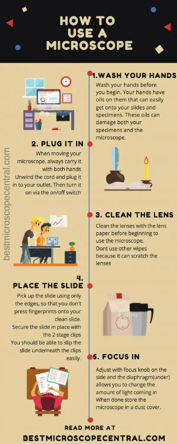
How to Use a Compound Microscope Basic Microscopy
How to Use a Compound Microscope Basic Microscopy. From the Virtual Microbiology Classroom on ScienceProfOnline.com. Image: The Far Side by Gary Larson. What am I going to learn from Lab Topic #1?. How to make a wet mount slide (Sweeeet) !

How to Use a Compound Microscope Basic Microscopy
E N D
Presentation Transcript
How to Use a Compound MicroscopeBasic Microscopy From the Virtual Microbiology Classroom on ScienceProfOnline.com Image: The Far Side by Gary Larson
What am I going to learn from Lab Topic #1? • How to make a wet mount slide(Sweeeet)! • How to microscopically view wet mounted plant cells & animal cells. • How to use and maintain a compound light microscope. • How to protect yourself from other people’s bodily fluids . From the Virtual Microbiology Classroom on ScienceProfOnline.com Image: Chimp brain in a jar, Gaetan Lee
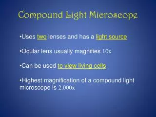
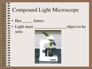
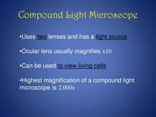
Compound Light Microscope The “Compound” Part • Simple microscopes have single magnifyinglens (like a magnifying glass). • Compound microscopes have two sets of lenses for magnification. • Lens closer to the eye = ocular lens (magnifying power of 10x). • Lenses closer to the object being viewed = objective lens. (Most light microscopes used in biology have three or four objective lenses). The “Light” Part • Bright-field light microscopes produce a dark image against brighter, backlit background. • Provide a 2-D image. • Commonly used to view stained cells. Image: Magnifying lamp use to look for part defects, US Navy; Compound light microscope, Moisey From the Virtual Microbiology Classroom on ScienceProfOnline.com
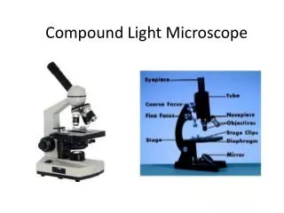
Your Microscope ARM Take Care of Your Scope: • It is your responsibility to take care of your scope and learn to use it properly. • I randomly check scopes to see if they are put away correctly. If yours is not, I may subtract points from your lab grade. Microscope Log: • If you get your scope out and find that it has not been put away properly, make an entry in the microscope log. Getting Scope Out: • The scope that you use is numbered to correspond to your Group number, as assigned by Mr. Banerjee. • When transporting your scope, always hold it with one hand under the base, and one hand around the arm. Putting Scope Away: • Clean stage if it is oily, and use lens paper to clean lenses. • Shortest objective lens (the one with the red band) should be pointing down toward stage. • Use course focus to position stage as low as it can go. • Always put scope back in numbered parking spot in scope cabinet. ** Now everyone get out their scope ** BASE From the Virtual Microbiology Classroom on ScienceProfOnline.com Image: Compound light microscope, Moisey
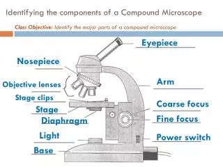
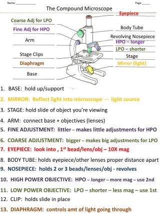
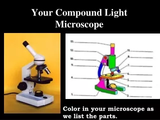
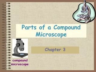
Parts of a Compound Light Microscope From the Virtual Microbiology Classroom on ScienceProfOnline.com Image: Compound light microscope, Moisey
Magnification & The Objective Lenses • Ocular lens magnifies the specimen 10x. • You will always be looking through the ocular and objective lens simultaneously, so multiply ocular magnification x objective power to calculate the Total Magnification (xTM). • Rotary nosepiece of your microscope has four objective lenses attached. • Shortest lens (red band) should have been pointing down when your scopes were last put away. From the Virtual Microbiology Classroom on ScienceProfOnline.com Image: Microscope objective lenses, T. Port
Scanning Power Objective Lens • Red band around it. • Magnifies objects 4x. • Q: What is the Total Magnification? ____ TM • We will only use this lens in today’s lab. It is not useful for looking at bacteria. From the Virtual Microbiology Classroom on ScienceProfOnline.com Image: Microscope objective lenses, T. Port
Low Power Objective Lens • Has yellow band around it. • Magnifies objects 10x. • Q: What is the Total Magnification? ____ TM • Start with this lens when looking at a bacterial smear. • Q: What does the term parfocal mean? From the Virtual Microbiology Classroom on ScienceProfOnline.com Image: Microscope objective lenses, T. Port
High Dry Objective Lens • Has blue band around it. • Magnifies objects 40x. • Q: What is the Total Magnification? ____ TM • Switch to this lens after getting your specimen in focus at 100xTM. From the Virtual Microbiology Classroom on ScienceProfOnline.com Image: Microscope objective lenses, T. Port
Microscopy:Electron Microscopes • Many microscopic images in your textbook were obtained using electron microscopes. • Electron beam wavelengths are shorter than light wavelengths, so better resolving power. • Q: What is resolution, in the context of microscopy? • Transmission Electron Microscope (TEM): 2-D image • Scanning Electron Microscope (SEM): 3-D image Images: Poliovirus, taken with TEM, PHIL #1875, Blood cells, taken with SEM, National Cancer Institute; Scanning electron microscope, Geological Survey of Israel laboratory. From the Virtual Microbiology Classroom on ScienceProfOnline.com
How to make a wet mount From the Virtual Microbiology Classroom on ScienceProfOnline.com Image: Wet mount procedure, T. Port
Procedure 1. How to make a wet mount 2. Letter “e” • What happens to the “e” when you look at it through the lens? • What happens to your view of the “e” when your increase magnification? 3. Onion • Wet mount, use stain • Note nucleus and cell wall • View video tutorial: 4. Geranium - Wet mount NO stain - Depth of field • 2 layers of cells • Note cell wall, chloroplasts streaming • View video tutorial: 5. Cheek cell • Wet mount using NaCl & Methylene blue. • Contrast! • View video tutorial: Images: Onion cells, Elodea cells & Cheek cells, SPO General Microbiology Microscopic Images; Elodea plant, Frank Vincentz From the Virtual Microbiology Classroom on ScienceProfOnline.com
Confused? Here are links to fun resources that further explain use of the microscope: • Microscopy Laboratory Main Pageon the Virtual Microbiology Classroom of Science Prof Online. • Compound Microscope Parts and Usevideofrom Science ProfOnline. • How to Make a Wet Mount of an Elodea Plant Cellvideo from ScienceProfOnline. • How to Make a Wet Mount of an Onion Epithelial Cellvideo from ScienceProfOnline. • How to Make a Wet Mount of a Cheek Cellvideo from ScienceProfOnline. • Play Amoeba, a video game where you are an amoeba that eats and grows. • Microscope Mania crossword puzzle. • Microscopic Pond Life, an extremely cool collection of videos of a variety of microscopic pond life to the tune of Radiohead’s “Kid A”. Smart Links From the Virtual Microbiology Classroom on ScienceProfOnline.com (You must be in PPT slideshow view to click on links.)
Are microbes intimidating you?Do yourself a favor. Use the…Virtual Microbiology Classroom (VMC) !The VMC is full of resources to help you succeed, including: practice test questions review questions study guides and learning objectives You can access the VMC by going to the Science Prof Online website www.ScienceProfOnline.com Images: White blood cell, Giant Microbes; Prokaryotic cell, Mariana Ruiz