Download
1 / 30
470 likes | 1.45k Views
Parts of a Compound Microscope. Chapter 3. Compound Microscope. A microscope is a very powerful magnifying glass A microscope helps you see things like cells up close. Eyepiece. View the specimen through the eyepiece. Stage Clips & Objectives. Stage clips hold the slide in place

E N D
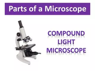
Parts of a Compound Microscope Chapter 3
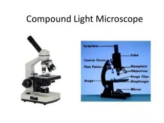
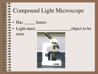
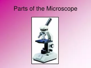
Compound Microscope • A microscope is a very powerful magnifying glass • A microscope helps you see things like cells up close
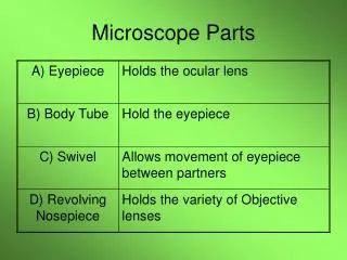
Eyepiece • View the specimen through the eyepiece
Stage Clips & Objectives • Stage clips hold the slide in place • Low power objective (the shorter one) is used to focus the microscope • High power objective is used to view fine details of a specimen
Coarse Adjustment,FineAdjustment, & Base • Coarse adjustment focuses first • Fine adjustment fine tunes & gives detailed focus (usually smaller than coarse adjustment knob) • Base is where the microscope rests
Stage • Stage is part where the slide rests • Mirror (or light source) directs light upwards onto the slide.
Diaphragm • Diaphragm allows light in
Nosepiece • Nosepiece is the rotating device that holds the objectives (lenses)
Arm • Arm is the part where you carry the microscope
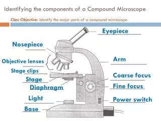
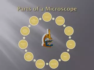
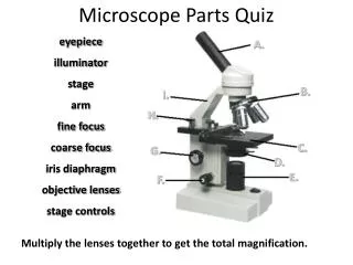
Answers • base • mirror (light source) • diaphragm • stage • stage clips • low power objective lens • high power objective lens • nosepiece • arm • fine focus knob • body tube • coarse focus knob • eyepiece
Identify the Parts of a Microscope Use Text pg. 688 to help you!
Body tube • Nosepiece • High power objective lens • Stage • Diaphragm • Eyepiece • Arm • Stage clip • Nothing • Coarse adjustment knob • Fine adjustment knob • Base • Low power objective lens • Medium power objective lens • Light source AnswersMake corrections
Main Types of Microscopes • Light microscopes bend light rays through lenses. • They are powerful because you multiply the powers of each lens to get total magnification 10x 20x What is the TOTAL MAGNIFICATION? 10 X 20 = 200x
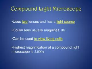
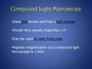
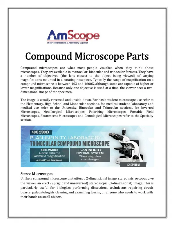
Types of Microscopes Compound Light Microscope • Has more than one lens in a tube • Shines beam of light through specimen • Specimens usually have to be stained with a chemical so they can be seen • Can magnify from 10x to 1000x
Electron Microscopes • Electron Microscopes use a focused beam of electrons instead of light • Increased magnification (from 100,000x to 1,000,000x) • Unfortunately it kills the specimen if it is alive.
The SEM shows very detailed 3-dimensional images at much higher magnifications than is possible with a light microscope. The images created without light waves are rendered black and white
Who am I? I’m a louse fly of a wallglider (an alpine bird)
Guess What? • You will have a QUIZ on these lecture notes (just microscope parts and functions!!!) on Friday, October 14th! Omnis cellula, e cellula
27 How Big is a Nanometer? • Consider a human hand skin white blood cell DNA atoms nanoscale Source: http://www.materialsworld.net/nclt/docs/Introduction%20to%20Nano%201-18-05.pdf
Sticky Spider Toes These are the single hairs (setae) that make up the tuft of hair on the bottom of a jumping spider’s foot. The oval represents the approximate size of the foot magnified to 270x. Water strider toes help keep it dry, but this spider’s toes help make him sticky! This picture, magnified 8750x, shows the very dense nanosized setules on the underside of just one of those many seta (hairs) shown in the picture above. http://www.primidi.com/2004/04/26.html Tell me more!
Lots of nano-toes! • Beetles and flies also have nanostructures that help them stick to walls, ceilings and what appear to be smooth surfaces.Tell me more! • http://shasta.mpi-stuttgart.mpg.de/biomaterials.html http://shasta.mpi-stuttgart.mpg.de/research/Bio-tribology.htm
For Monday’s Lab . . . • Bring in some printed materials to look at in our lab (newspaper, magazines, comics, catalogs, stamps, etc.) • Other good items to bring: pet hair, feathers, scales (put in a baggie and label so we know what it is!)