Download
1 / 40
560 likes | 850 Views
RADIOLOGY ANATOMY OF THE PITUITARY GLAND. Dr. fahad albadr. ANATOMY OF THE PITUITARY GLAND. OBJECTIVES. At the end of the lecture, students should be able to: Describe the position of the pituitary gland. List the structures related to the pituitary gland.

E N D
RADIOLOGY ANATOMY OF THE PITUITARY GLAND Dr. fahad albadr
OBJECTIVES At the end of the lecture, students should be able to: • Describe the positionof the pituitary gland. • List the structures relatedto the pituitary gland. • Differentiate between the lobes of the gland.
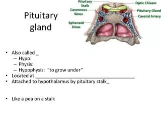
PITUITARY GLAND(HYPOPHYSIS CEREBRI) It is referred to as the master of endocrine glands. It is a small oval structure 1 cm in diameter.
X-RAY SKULL: LATERAL VIEW SAGITTAL SECTION OF HEAD & NECK PITUITARY GLAND Pituitary gland Hypophyseal fossa Sphenoidal air sinus
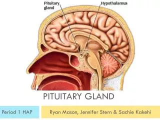
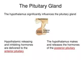
It lies in the middle cranial fossa It is well protected in sellaturcica (hypophysealfossa) of body of sphenoid POSITION Sella turcica
Optic chiasma Mamillary body Body of sphenoid it lies between optic chiasma(anteriorly) & mamillary bodies (posteriorly).
A fold of dura mater (Diaphragma sellae) covers the pituitary gland & has an opening for passage of infundibulum (pituitary stalk) connecting the gland to hypothalamus.
IMPORTANT RELATIONS SUPERIOR: Diaphragmasellae INFERIOR: Sphenoidal air sinuses LATERAL:Cavernous sinuses
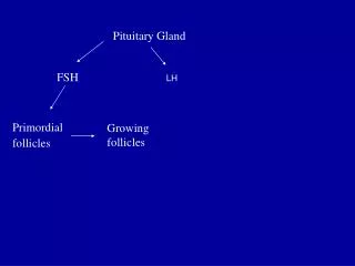
Hypothalamo-hypophyseal tract SUBDIVISIONS OF PITUITARY GLAND The gland is subdivided into: 1) Anterior lobe (Adenohypophysis): it is the True gland, Secretes hormones 2) Posterior lobe (Neurohypophysis): connected to hypothalamus through hypothalamo-hypophyseal tract, Stores hormones secreted by hypothalamic nuclei
BLOOD SUPPLY OF PITUITARY GLAND ARTERIES: Superior & inferior hypophyseal arteries (branches of internal carotid artery) VEINS:Hypophyseal veins drain into Cavernous Sinuses.
DISTRIBUTION OF ARTERIES a hypothalamo- hypophseal portal vessel • Superior hypophyseal: supplies infundibulum & forms a capillary network from which vessels pass downward & form sinusoids into the anterior lobe of pituitary gland (hypophyseal portal system). • Inferior hypophyseal: supplies posterior lobe of pituitary gland.
ANTERIOR LOBE Hormone-releasing & inhibiting factors produced by hypothalamus use Hypophyseal Portal Systemof vessels to reach the Anterior lobe of pituitary gland
POSTERIOR LOBE The neurohypophysis receives a nerve supply from some of the hypothalamic nuclei (supraoptic& paraventricular) The axons of these nuclei convey their neurosecretionto the Posterior lobe of pituitary gland through Hypothalamo-Hypophyseal tract from where it passes into the blood stream.
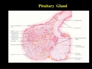
NORMAL PITUITARY GLAND • The gland is composed of two parts: • Anterior lobe (adeno hypophysis) • Posterior lobe (neuro hypophysis) • Normal size: • Weight: 0.5g • Height: 4-12 mm • Anterior posterior: 5-16 mm
INDICATIONS FOR IMAGING THE PITUITARY GLAND • Hormonal dysfunction • Cushing syndrome • Growth abnormalities e.g. Growth hormone deficiency, acromegaly • Visual abnormalities • headache
What is best modality to image the pituitary gland ? • X ray • CT scan • MRI • US • Nuclear medicine
What is best modality to image the pituitary gland ? • X ray • CT scan • MRI • US • Nuclear medicine
CT scan MRI
CT scan MRI
1 2 3 4 5 6
1 2 1-Optic sulcus 2- Anterior clinoid process 3-Floor of sella turcia (Pituitary fossa) 4- Posterior clinoid process 5- Dorsum sella 6- Sphenoid sinus 3 4 5 6
4 3 5 2 6 1
4 3 1- pituitary gland 2- sphenoid sinus 3- optic chiasm 4- hypothalamus 5- pituitary stalk 6- claivus 5 2 6 1
NORMAL PITUITARY ADENOMA
2 3 1 4 5 6
2 3 1 4 5 6
Optic chiasm Pituitary stalk Pituitary gland Carotid artery Cavernous sinus Sphenoid sinus