Download
1 / 19
190 likes | 212 Views
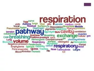
Explore the mechanics of breathing in the respiratory system, from inhalation to exhalation. Learn about the bronchial tree, gas exchange, involuntary respiration, and lung capacities. Understand how the diaphragm and intercostal muscles work. Discover terms like tidal volume, inspiratory reserve volume, and more.

E N D
Ventilation: The Mechanics of Breathing Miss Ulrich
Lungs close up… • Bronchial Tree Consisting of the Passageways that Connect the Trachea and Alveoli Q: Where does gas exchange occur?
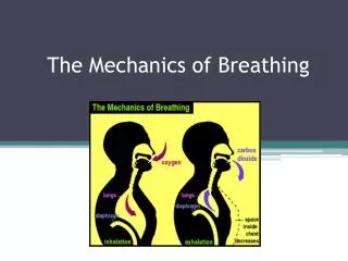
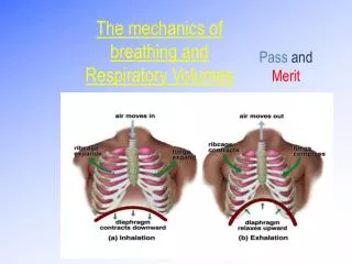
Breathing • The movement of air into and out of the lungs (ventilation) results from a pressure difference between the thoracic cavity and the atmosphere. • This pressure difference is created by changing the volume of the thoracic cavity.
Involuntary Respiration. The basic rhythm of breathing occurs without conscious effort. The inspiratory center located in the medulla sets the basic rhythm by automatically initiating inspiration with a two second burst of nerve impulses to the diaphragm and the external intercostal muscles. Contraction of the diaphragm and the external intercostal muscles draws air into the lungs. Involuntary Respiration
The Expiratory Center. The expiratory center is located in the medulla. This center functions during forced expiration stimulating the internal intercostal and abdominal muscles to contract.
Inhalation • During inhalation, the diaphragm contracts and flattens and the external intercostal muscles draw the ribs upward and outward. • This increase in thoracic volume results in a decrease in intrapulmonary pressure. • Air enters the lungs to stabilize the pressure difference between the external atmosphere and the internal compartments of the lungs. • Normal inhalation is an active process, requiring muscular work.
During quiet breathing, intercostals maintain the rigidity of the chest wall. Otherwise, reduced intra-thoracic pressure would cause the chest wall to collapse inwards. External Intercostals (on the outside of the ribcase) wrap around from the back of the rib almost to the end of the bony part of the rib in front. . They elevate the ribs. Internal Intercostals in the inside of the ribcase) extend from the front of the ribs, and go around back, past the bend in the ribs . They depress the ribs.
Diaphragm • In a healthy adult, the diaphragm is the dominant muscle of respiration at rest • The diaphragm is a musculotendinous sheet separating the thorax from the abdomen. It is attached to the thoracic cage under the lower ribs.
Expiration • Exhalation is normally a passive process. • The diaphragm and external intercostal muscles relax decreasing the volume of the thoracic cavity. • This causes the pressure within the lungs to exceed the atmospheric pressure. • Air is expelled from the lungs.
Forced Exhalation • During a forced exhalation, the internal intercostal muscles contract, depressing the rib cage. • The abdominal muscles contract, pushing the organs in the abdominal cavity against the diaphragm. • The thoracic volume decreases to a level lower than achieved in normal exhalation. • These muscles are used to counteract the effects of obstructive pulmonary disorders.
ERV • These muscles are used during a forced exhalation to determine the expiratory reserve volume (ERV). • ERV is - the maximum volume of gas that can be forcefully exhaled after a normal exhalation (tidal volume).
The volume of the lungs is divided into four functional compartments, lung volumes. Combinations of two or more lung volumes are called a lung capacity. Other Terms of Breathing
Terms • tidal volume ( TV ) - the volume of gas inspired or expired during each normal (unforced) ventilation cycle (volume of air moved into the lungs in a single breath. • inspiratory reserve volume ( IRV ) -the maximum amount of gas that can be forcefully inhaled after a normal inhalation. • expiratory reserve volume ( ERV ) - the maximum volume of gas that can be forcefully exhaled after a normal exhalation.
Terms • residual volume ( RV ) - the amount of gas left in the lungs after a maximum (forced) exhalation. Necessary otherwise the lungs would collapse. • total lung capacity ( TLC ) - the amount of gas in the lungs after a maximum (forced) inhalation. TLC = IRV + TV + ERV + RV • vital capacity ( VC )-the maximum volume of gas that can be exhaled by voluntary effort after a maximum inhalation. VC = IRV + TV + ERV
Terms • inspiratory capacity ( IC ) - the maximum amount of gas that can be inhaled after a normal (unforced) exhalation. IC = IRV + TV • functional residual capacity ( FRC ) - the amount of gas left in the lungs after a normal (unforced) exhalation. FRC = ERV + RV