Download
1 / 20
200 likes | 328 Views
This study investigates the structural and electronic properties of nitrosoiron(II) porphyrins using electron paramagnetic resonance (EPR) g-tensors derived from density functional theory (DFT). We reveal insights into the coordination of nitric oxide around iron in heme nitrosyls, including the characterization of distinct radical species. Our findings propose a new structural model for axial species and clarify contributions to g-tensor deviations from the free electron value. This work enhances understanding of iron-containing porphyrin complexes in biological systems.

E N D
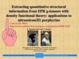
Extracting quantitative structural information from EPR g-tensors with density functional theory: applications to nitrosoiron(II) porphyrins I am on the Web: http://www.cobalt.chem.ucalgary.ca/ps/posters/EPR-FeP/ S. Patchkovskii and T. Ziegler Department of Chemistry, University of Calgary, 2500 University Dr. NW, Calgary, Alberta, T2N 1N4 Canada Structure of heme nitrosyls from EPR g-tensors with DFT CSC’83, 2000
Introduction. Conclusions and outlook Introduction Iron-containing porphyrin complexes (hemes) serve as prosthetic groups in many vitally important enzymes[1,2], such as hemoglobin. Because oxy-hemoglobin possesses a singlet electronic structure, it is not amenable to studies using electron paramagnetic resonance (EPR). Structurally similar heme nitrosyls have a spatially non-degenerate spin-doublet ground state, which is readily observable with EPR. Experimental EPR spectra of nitrosylated hemoglobins and myoglobins[3,4], indicate the presence of two distinct radical species: rhombic and axial. The rhombic ("Type I") species exhibits three distinct principal components in its EPR g tensor (g1=1.96-1.98, g2=2.00, g3=2.06-2.08). It has been assigned to a six-coordinated structure, with NO coordinated in a bent end-on orientation, and the second axial position filled by an imidazole side chain. Despite extensive investigations, the nature of the axial ("Type II") species (g||=1.99-2.00, g=2.02-2.03) have proven more elusive, and remains controversial. In this work, we examine the structure and EPR g-tensors of model porphyrins with density functional theory (DFT). On the basis of our calculations, we propose a new structural model for the axial species. Structure of heme nitrosyls from EPR g-tensors with DFT CSC’83, 2000
Theory I Theory Quasi-relativistic DFT formulation of the EPR g-tensors used in this work distinguishes between several contributions to the g-tensor[5,6]: free-electron g value (2.0023) diamagnetic term paramagnetic term Darwin term Relativistic corrections mass-velocity correction kinetic energy correction The paramagnetic term dominates deviation of g from the free-electron value for complexes considered here, and can be in turn separated into several contributions: frozen core contributions occupied-virtual coupling terms occupied-occupied coupling terms The occ-vir term is usually the most qualitatively important contribution. Structure of heme nitrosyls from EPR g-tensors with DFT CSC’83, 2000
Theory II The contribution is given by (atomic units): 0.00731 -spin current the effective potential -spin current due to unit magnetic field along s=x,y, or z The form of the occupied-virtual paramagnetic contribution the the EPR g-tensor is analogous to the expression for the paramagnetic part of the NMR shielding tensor for a nucleus N, given by: The similarity between the two quantities is extremely useful both in the evaluation and in analysis of g tensor, and is unique to our DFT implementation. Structure of heme nitrosyls from EPR g-tensors with DFT CSC’83, 2000
Theory III The spin-current density for a spin arising due to the coupling between occupied and virtual MOs caused by the external magnetic field B0 is given by: magnetic coupling coefficient field strength in the direction s (=x,y,z) unperturbed occupied MO unperturbed virtual MO The principal contribution to the coupling coefficient u is in turn given by: atomic orbitals (AOs) applies to each AO unperturbed orbital energies unperturbed MO coefficients Structure of heme nitrosyls from EPR g-tensors with DFT CSC’83, 2000
Methods Methods Structure of heme nitrosyls from EPR g-tensors with DFT CSC’83, 2000
ON-Fe(P): Coordination of NO Conclusions and outlook 1a: +3.9 kcal/mol 1b: +0.0 kcal/mol 1c: +0.0 kcal/mol ON-Fe(P): Coordination of NO Z • The coordination of NO around iron gives rise to three key points on the PES of 1, namely: • the C4v structure 1a with the linearly coordinated NO ligand.This is a second-order saddle point, rather than a minimum. • Cs structure 1b, with bent end-on coordination of the NO ligand, pointing towards one of the meso carbon atoms of the porphyrin ligand. • a second Cs structure 1c, with the NO bond eclipsing one of the equatorial Fe-Np bonds. • Given the isoenergetic 1a and 1b, the NO ligand in free five-coordinated heme nitrosyl should undergo free intramolecular rotation around the Z axis. A closer inspection of the optimised structures reveals that the rotation of the NO ligand is coupled to the distortion of the porphyrin ligand. As a consequence, hindering the distortion increases the barrier for NO rotation increases to about 1.2 kcal/mol. Structure of heme nitrosyls from EPR g-tensors with DFT CSC’83, 2000
ON-Fe(P)-Im: Coordination of imidazole Conclusions and outlook 3.0 E, kcal/mol 2.0 1.0 RFe-N(Im),Å 2.0 2.2 2.4 2.6 2.8 ON-Fe(P)-Im: Coordination of imidazole Dissociation of the imidazole was examined by gradually increasing the iron-imidazole distance, and optimizing the rest of the structure. The resulting energy profile is exceptionally flat, with variations in the Fe-N(Im) bond length in the 2.05-2.50Å range changing energy by less than 1 kcal/mol. As a consequence, experimental bond lengths are likely to be influenced by substitution and environment effects. Although the potential energy profile for the dissociation of imidazole appears to indicate a presence of a local minimum at 2.4Å, g-tensors show no qualitative changes in the vicinity of this structure. exp Structure of heme nitrosyls from EPR g-tensors with DFT CSC’83, 2000
ON-Fe(P)-Im: rotation of the axial ligands Conclusions and outlook 2e 2a 2d 2b 2c 0.60 energy, kcal/mol 0.40 0.20 Im= 0º NO, degree Im=45º 0.00 0 50 100 150 200 250 300 350 In the six-coordinated complex, both axial ligands preferentially appear in staggered orientation (2a-2c). The local minima appear within 0.3 kcal/mol of the global minimum (2c), and will be substantially populated at non-zero temperature. The orientations of NO and imidazole in condensed phases are likely to be determined by substituent effects and intermolecular interactions. Free rotation of both axial substituents may also be expected at room temperature. Structure of heme nitrosyls from EPR g-tensors with DFT CSC’83, 2000 32
Origin of g in C4v structure. Conclusions and outlook eV +3.0 +2.0 1* +1.0 SOMO -36 -spin 0.0 -1.0 -spin n1 +20 -2.0 2 Contributions to g are in parts per thousand (ppt) -3.0 +14 1 -4.0 Origin of the g tensors: C4v structure Mulliken d-population on iron in 1 (6.7) is consistent with d7 electron count, formally making 1 a complex of FeI and NO+. The unpaired electron is mostly on iron’s dz2 AO. The SOMO vanishes upon action of the Mz operator, so that all spin-restricted terms in the parallel component g|| also vanish. If the magnetic field is in the XY plane, the field-induced coupling with -spin * MOs (dxz, dyz-like), localised on the Fe-N=O fragment, results in a contribution of -36 ppt to g. This contribution is almost identically cancelled by two -spin terms, (+14 and +20 ppt) Structure of heme nitrosyls from EPR g-tensors with DFT CSC’83, 2000
Origin of g in meso-Cs structure Conclusions and outlook eV +3.0 +2.0 1* +1.0 -13 -15 0.0 +20 -1.0 -36 +32 n1 -2.0 +37 2 -3.0 +7 +14 1 -4.0 bent linear Origin of the g tensors: Cs structures In the bent 1b, iron's dz2 AOs overlap with the * orbitals of the NO ligand, tilting the dz2-like contribution to the SOMO towards the direction perpendicular to the N=O bond. Such orientation allows SOMO to interact with occupied non-bonding dx2-y2 orbital n1. The SOMO and SOMOn terms exhibit a qualitatively different dependence on the relative orientation of the NO ligand and the porphyrin core, so that g-tensor in bent 1 depends on the orientation of NO. Structure of heme nitrosyls from EPR g-tensors with DFT CSC’83, 2000
Origin of g in ON-Fe(P)-Im Conclusions and outlook Origin of the g tensors: ON-Fe(P)-Im 1b: SOMO 2b: SOMO Upon coordination of the imidazole, a weak * interaction between an imidazole lone pair, and the dz2-like lobe of the SOMO, destabilises the SOMO by 0.9 eV. The Mulliken spin population on iron is also reduced, from 0.9 (1b) to 0.5 (2b). • decrease in the d character of the SOMO reduces all spin-orbit coupling matrix elements, decreasing all g’s. • a large NO * contribution to the SOMO increases the magnitude of matrix elements of the M operator in -SOMO - * terms. • destabilisation of the SOMO increases -SOMO - * terms, while decreasing -SOMO - contributions. Structure of heme nitrosyls from EPR g-tensors with DFT CSC’83, 2000
Numerical results: Principal components Conclusions and outlook Numerical results: Principal components As expected from the qualitative analysis, calculated principal g-tensor components in ON-Fe(P) (1) are sensitive to both the Fe-N=O bond angle, and to the orientation of the NO relative to the porphyrin (1a-1c). The values are only in a broad qualitative agreement with experiment, and appear to indicate an orientation of the NO ligand intermediate between 1b and 1c.Part of the error may be due to the neglect of the environment effects. In ON-Fe(P)-Im (2), g-tensor is again sensitive to the orientation of NO, but is left unchanged by imidazole rotation. Calculated magnitudes of the principal components appear to be in a broad qualitative agreement with experiment. The characteristic g1<g2(ge)<<g3 pattern is reasonably well reproduced. At the same time, calculated values are too small compared to experiment, by 20 to 50 ppt. *: TPP = tetraphenylporphyrinato2-; Mb = myoglobin; Hb = hemoglobin Structure of heme nitrosyls from EPR g-tensors with DFT CSC’83, 2000
Numerical results: g-tensor orientations Conclusions and outlook gfree g2 g1 g1 g3 g2 Numerical results: g tensor orientation Theoretical orientations of the g-tensor components in 2 are in a good agreement with experiment for low-temperature "Type I" nitroso myoglobin. At 77K, the principal components of the g tensor in MbNO[3] deviate from the direction perpendicular to porphyrin plane (Z axis) by, respectively, 63±2º (g1), 27±2º (g2), and 88±2º (g3). The corresponding theoretical values for free 2a are 58º, 32º, and 90º. Comparison of the remaining experimental direction cosines with the theoretical results gives of approximately -70º. The good agreement in the theoretical and experimental orientations of the g-tensor indicated that the {FeNO} fragment in MbNO is not substantially distorted by the protein. The large g2 orientation has been taken previously[4] as an indication of the Fe-N(NO) axial bond direction. As can be seen from the SOMO plots, it in fact corresponds to the direction of the dz2-like part of the SOMO, which in turn deviates from the Fe-N(NO) bond by almost 40º. Structure of heme nitrosyls from EPR g-tensors with DFT CSC’83, 2000
Numerical results: RFe-N(Im) and g-tensor Conclusions and outlook 2.08 3.0 g3 E, kcal/mol 2.04 g g tensor components 2.0 g2 2.00 1.0 g||. g1 1.96 RFe-N(Im),Å 2.0 2.2 2.4 2.6 2.8 Numerical results: RFe-N(Im) and g-tensor Elongation of the Fe-N(Im) bond results in an increase in all three principal components. The free electron-like component (g2) increases by only 8 ppt/Å in the 2.0-2.9 Å range. The other two components, g1 and g3, increase at the rates of, respectively, 45 and 36 ppt/Å. At the separation of 2.9 Å or more, the calculated g tensor becomes essentially identical to the result for free five-coordinated species. If the NO ligand rotates freely on the EPR time scale, averaging results in g> g||. The parallel component increases the rate of 19 ppt/Å. g grows at 35 ppt/Å, so that the separation between the rotationally averaged components increases with dissociation of imidazole. g tensor components Structure of heme nitrosyls from EPR g-tensors with DFT CSC’83, 2000
Biological ON-Fe(P)-Im: Models galore Conclusions and outlook Biological ON-Fe(P)-Im: Models galore P.E.S. g-tensor Model Rhombic Axial Source Axial structure is not a minimum; axial g-tensor components are wrong linear- bent [3] “Axial” structure is not a minimum, and has rhombic g-tensor up- down [13] Rhombic structure is not a minimum; “Axial” structure has rhombic g-tensor. inclined- straight [15] bound- dissociated “Axial” structure has rhombic g-tensor. [16] Structure of heme nitrosyls from EPR g-tensors with DFT CSC’83, 2000
Biological ON-Fe(P)-Im: rhombic species Conclusions and outlook Biological ON-Fe(P)-Im: Rhombic species The rhombic signal ("State I") exhibits a characteristic pattern of gmin<gfree<<gmax. This pattern appears to correspond to the six-coordinated complex 2 with the internal rotation of the NO ligand frozen on the EPR time scale. Calculated orientations of the principal components are in an excellent agreement with the experimental result for nitrosylated myoglobin (MbNO) single crystals. At low temperatures, the direction of the g2 component in MbNO deviates by 30º from the average normal to the porphyrin plane. The value calculated at the theoretical gas-phase geometry of the model complex 2 is 32º, suggesting that the ON-Fe(P) fragment in MbNO is not noticeably distorted by interactions with the protein environment. Structure of heme nitrosyls from EPR g-tensors with DFT CSC’83, 2000
Biological ON-Fe(P)-Im: axial species Conclusions and outlook Biological ON-Fe(P)-Im: Axial species The axial (“Type II”) spectra are usually found at higher temperatures. In the absence of a satisfactory static model, it appears reasonable to consider possible dynamical interpretations. If the rotation of NO is unfreezed, averaging of the g components leads to an axial spectrum. In the model complex, imidazole has no additional chemical bonds to the ON-Fe(P) fragment, and will dissociate completely. In heme proteins, it is provided by a histidine residue, and is tethered to the backbone. The tethering may prevent a complete dissociation, leading to smaller values of g and g||. It also allows protein to switch between Type I and Type II spectra at the same temperature: A conformational change in the backbone can pull the imidazole from the rest of the complex, effectively causing its dissociation. This would, in turn, lower the barrier for NO rotation, and produce the change in the spectrum. Structure of heme nitrosyls from EPR g-tensors with DFT CSC’83, 2000
Summary Conclusions and outlook Summary Structure of ON-Fe(P)-Im: • In both five- and six-coordinated complexes, NO is preferably coordinated end-on, with a Fe-N=O bond angle of approximately 140º. • In the gas-phase five-coordinated complex, NO undergoes free rotation. Imidazole coordination in the second axial position increases the activation barrier • Variations in Fe-N(Im) bond in the 2.05-2.5Å change energy by less than 1 kcal/mol. EPR g-tensors: • are sensitive to the orientation of NO and Fe-N(Im) bond length. • are not sensitive to the orientation of the imidazole ligand • orientation of the g-tensor component, showing the smallest deviation from the free-electron value, does not coincide with the direction of the axial Fe-N(NO) bond. Biological ON-Fe(P)-Im systems: • Rhombic ("Type I") EPR signal in nitrosoheme systems corresponds to a static structure with NO in a staggered orientation, and RFe-N(Im)2.1Å. The ON-Fe(P) moiety is not noticeably distorted by the protein environment. • The axial ("Type II") EPR signal is tentatively assigned to a partially dissociated species (RFe-N(Im)2.5Å), with a freely rotating NO ligand. Structure of heme nitrosyls from EPR g-tensors with DFT CSC’83, 2000
Acknowledgements Acknowledgements Acknowledgements This work has been supported by the National Sciences and Engineering Research Council of Canada (NSERC), as well as by the donors of the Petroleum Research Fund, administered by the American Chemical Society (ACS-PRF No 31205-AC3). Dr. Georg Schreckenbach is gratefully acknowledged for making the GIAO-DFT implementation of the EPR g tensors available to the authors. References Structure of heme nitrosyls from EPR g-tensors with DFT CSC’83, 2000