Download

1 / 36
370 likes | 446 Views
Unveil the intricate workings of hormones and the endocrine system in maintaining homeostasis, including hormone types, functions, and mechanisms of action. Explore the dynamic interplay between hormones and target cells, eliciting precise regulatory responses.

E N D
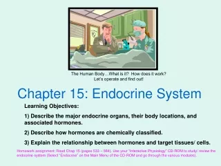
Introduction • The endocrine and nervous systems function to achieve and maintain homeostasis (Table 16-1) • When the two systems work together as one system, referred to as the neuroendocrine system, they perform the same general functions: communication, integration, and control • In the endocrine system, secreting cells send hormone molecules via the blood to specific target cells contained in target tissues or target organs
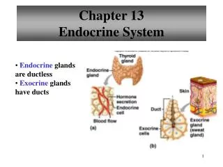
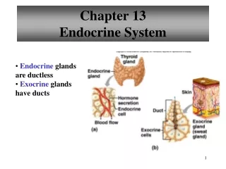
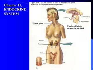
Introduction • Hormones—carried to almost every point in the body; can regulate most cells; effects work more slowly and last longer than those of neurotransmitters • Endocrine glands are “ductless glands”; many are made of glandular epithelium whose cells manufacture and secrete hormones; a few endocrine glands are made of neurosecretory tissue • Glands of the endocrine system are widely scattered throughout the body (Figure 16-2; Table 16-2)
Hormones • Classification of hormones • Classification by general function • Tropic hormones—target other endocrine glands and stimulate their growth and secretion • Sex hormones—target reproductive tissues • Anabolic hormones—stimulate anabolism in target cells • Classification by chemical structure (Figure 16-3; Table 16-3) • Steroid hormones • Nonsteroid hormones
Hormones • Classification of hormones (cont.) • Steroid hormones (Figure 16-4) • Synthesized from cholesterol (Figure 16-5) • Lipid-soluble and can easily pass through the phospholipid plasma membrane of target cells • Examples of steroid hormones: cortisol, aldosterone, estrogen, progesterone, and testosterone
Hormones • Classification of hormones (cont.) • Nonsteroid hormones (Figure 16-6) • Synthesized primarily from amino acids • Protein hormones—long, folded chains of amino acids; e.g., insulin and parathyroid hormone • Glycoprotein hormones—protein hormones with carbohydrate groups attached to the amino acid chain • Peptide hormones—smaller than protein hormones; short chain of amino acids; e.g., oxytocin and antidiuretic hormone (ADH) • Amino acid derivative hormones—each is derived from a single amino acid molecule • Amine hormones—synthesized by modifying a single molecule of tyrosine; produced by neurosecretory cells and by neurons; e.g., epinephrine and norepinephrine • Amino acid derivatives produced by the thyroid gland; synthesized by adding iodine to tyrosine
Hormones • How hormones work • General principles of hormone action • Hormones signal a cell by binding to the target cell’s specific receptors in a “lock-and-key” mechanism (Figure 16-7) • Different hormone-receptor interactions produce different regulatory changes within the target cell through chemical reactions • Combined hormone actions: • Synergism—combinations of hormones acting together have a greater effect on a target cell than the sum of the effects that each would have if acting alone • Permissiveness—when a small amount of one hormone permits, or enables,a second one to have its full effects on a target cell • Antagonism—one hormone produces the opposite effects of another hormone; used to “fine tune” the activity of target cells with great accuracy • Most hormones have primary effects that directly regulate target cells and many secondary effects that influence or modulate other regulatory mechanisms in target cells • Endocrine glands produce more hormone molecules than are actually needed; the unused hormones are quickly excreted by the kidneys or broken down by metabolic processes
Hormones • How hormones work (cont.) • Mechanism of steroid hormone action (Figure 16-8) • Steroid hormones are lipid-soluble, and their receptors are normally found in the target cell’s cytosol • After a steroid hormone molecule has diffused into the target cell, it binds to a receptor molecule to form a hormone-receptor complex • Mobile-receptor model—hormone passes into nucleus, where it binds to mobile receptor and activates a certain gene sequence to begin transcription of mRNA; newly formed mRNA molecules move into the cytosol, associate with ribosomes, and begin synthesizing protein molecules that produce the effects of the hormone • Steroid hormones regulate cells by regulating production of certain critical proteins • The amount of steroid hormone present determines magnitude of a target cell’s response • Because transcription and protein synthesis take time, responses to steroid hormones are often slow
Hormones • How hormones work (cont.) • Mechanisms of nonsteroid hormone action • The second messenger mechanism—also known as the fixed-membrane-receptor model (Figure 16-9) • A nonsteroid hormone molecule acts as a “first messenger” and delivers its chemical message to fixed receptors in the target cell’s plasma membrane • The “message” is then passed by way of a G protein into the cell, where a “second messenger” triggers the appropriate cellular changes • Second messenger mechanism—produces target cell effects that differ from steroid hormone effects in several important ways: • Effects of the hormone are amplified by the cascade of reactions • There are a variety of second messenger mechanisms—examples: IP3, GMP, calcium-calmodulin mechanisms (Figure 16-10) • The second messenger mechanism operates much more quickly than the steroid mechanism • The nuclear receptor mechanism—small iodinated amino acids (T4 and T3) enter the target cell and bind to receptors associated with a DNA molecule in the nucleus; this binding triggers transcription of mRNA and synthesis of new enzymes
Hormones • Regulation of hormone secretion • Control of hormonal secretion is usually part of a negative feedback loop and is called endocrine reflexes (Figure 16-11) • Simplest mechanism—when an endocrine gland is sensitive to the physiological changes produced by its target cells • Endocrine gland secretion may also be regulated by a hormone produced by another gland • Endocrine gland secretions may be influenced by nervous system input; this fact emphasizes the close functional relationship between the two systems
Hormones • Regulation of target cell sensitivity • Sensitivity of target cell depends in part on number of receptors (Figure 16-12) • Up-regulation—increased number of hormone receptors increases sensitivity • Down-regulation—decreased number of hormone receptors decreases sensitivity • Sensitivity of target cell may also be regulated by factors that affect signal transcription or gene transcription
Prostaglandins (PGs) • Unique group of lipid hormones (20-carbon fatty acid with 5-carbon ring) that serve important and widespread integrative functions in the body but do not meet the usual definition of a hormone (Figure 16-13; Table 16-4) • Called tissue hormones because the secretion is produced in a tissue and diffuses only a short distance to other cells within the same tissue; PGs tend to integrate activities of neighboring cells
Prostaglandins • Many structural classes of prostaglandins have been isolated and identified: • Prostaglandin A (PGA)—intraarterial infusion resulting in an immediate fall in blood pressure accompanied by an increase in regional blood flow to several areas • Prostaglandin E (PGE)—vascular effects: regulation of red blood cell deformability and platelet aggregation; inflammation (which can be blocked with drugs that inhibit PG-producing enzymes such as COX-1 and COX-2), gastrointestinal effects: regulates hydrochloric acid secretion • Prostaglandin F (PGF)—especially important in reproductive system, causing uterine contractions; also affects intestinal motility and is required for normal peristalsis • Many tissues are known to secrete PGs • PGs have diverse physiological effects
Pituitary Gland • Structure of the pituitary gland • Formerly known as hypophysis • Size: 1.2 to 1.5 cm (about 1⁄2 inch) across; weight: 0.5 g (1⁄60 ounce) • Located on the ventral surface of the brain within the skull (Figure 16-14) • Infundibulum—stemlike stalk that connects pituitary to the hypothalamus • Made up of two separate glands, the adenohypophysis (anterior pituitary gland) and the neurohypophysis (posterior pituitary gland)
Pituitary Gland • Adenohypophysis (anterior pituitary) • Divided into two parts: • Pars anterior—forms the major portion of adenohypophysis • Pars intermedia • Tissue is composed of irregular clumps of secretory cells supported by fine connective tissue fibers and surrounded by a rich vascular network • Three types of cells can be identified according to their affinity for certain stains (Figure 16-15): • Chromophobes—do not stain • Acidophils—stain with acid stains • Basophils—stain with basic stains
Pituitary Gland • Adenohypophysis (anterior pituitary) (cont.) • Five functional types of secretory cells exist: • Somatotrophs—secrete GH • Corticotrophs—secrete ACTH • Thyrotrophs—secrete TSH • Lactotrophs—secrete prolactin (PRL) • Gonadotrophs—secrete LH and FSH
Pituitary Gland • Adenohypophysis (anterior pituitary) (cont.) • Growth hormone (GH) (Figure 16-16; Table 16-6) • Also known as somatotropin (STH) • Promotes growth of bone, muscle, and other tissues by accelerating amino acid transport into the cells • Stimulates fat metabolism by mobilizing lipids from storage in adipose cells and speeding up catabolism of the lipids after they have entered another cell • GH tends to shift cell chemistry away from glucose catabolism and toward lipid catabolism as an energy source; this leads to increased blood glucose levels • GH functions as an insulin antagonist and is vital to maintaining homeostasis of blood glucose levels
Pituitary Gland • Adenohypophysis (anterior pituitary) (cont.) • Prolactin (PRL) (Table 16-6) • Produced by acidophils in the pars anterior • Also known as lactogenic hormone • During pregnancy, PRL promotes development of the breasts, anticipating milk secretion; after the baby is born, PRL stimulates the mother’s mammary glands to produce milk
Pituitary Gland • Adenohypophysis (anterior pituitary) (cont.) • Tropic hormones—have a stimulating effect on other endocrine glands; four principal tropic hormones are produced and secreted by the basophils of the pars anterior (Table 16-6): • Thyroid-stimulating hormone (TSH), or thyrotropin—promotes and maintains growth and development of thyroid; also causes thyroid to secrete its hormones • Adrenocorticotropic hormone (ACTH), or adrenocorticotropin—promotes and maintains normal growth and development of cortex of adrenal gland; also stimulates adrenal cortex to secrete some of its hormones • Follicle-stimulating hormone (FSH)—in female, stimulates primary graafian follicles to grow toward maturity; also stimulates follicle cells to secrete estrogens; in male, FSH stimulates development of seminiferous tubules of testes and maintains spermatogenesis • Luteinizing hormone (LH)—in female, stimulates formation and activity of corpus luteum of ovary; corpus luteum secretes progesterone and estrogens when stimulated by LH; LH also supports FSH in stimulating maturation of follicles; in male, LH stimulates interstitial cells in testes to develop and secrete testosterone; FSH and LH are called gonadotropins because they stimulate growth and maintenance of gonads
Pituitary Gland • Adenohypophysis (anterior pituitary) (cont.) • Control of secretion in the adenohypophysis • Hypothalamus secretes releasing hormones into the blood, which are then carried to hypophyseal portal system (Figure 16-17; Table 16-5) • Hypophyseal portal system carries blood from hypothalamus directly to adenohypophysis where target cells of releasing hormones are located (Figure 16-18) • Releasing hormones influence secretion of hormones by acidophils and basophils • Through negative feedback, hypothalamus adjusts secretions of adenohypophysis, which then adjusts secretions of target glands that, in turn, adjust activity of their target tissues (Figure 16-19) • Minute-by-minute variations in hormone secretion can exhibit occasional large peaks, caused by pulse in releasing hormone secretion by hypothalamus (Figure 16-20) • In stress, hypothalamus translates nerve impulses into hormone secretions by endocrine glands, basically creating a mind-body link
Adrenal Glands • Adrenal cortex (cont.) • Mineralocorticoids • Have an important role in regulatory process of sodium in the body • Aldosterone • Only physiologically important mineralocorticoid in the human; primary function is maintenance of sodium homeostasis in the blood by increasing sodium reabsorption in the kidneys • Aldosterone also increases water retention and promotes loss of potassium and hydrogen ions • Aldosterone secretion is controlled by the renin-angiotensin-aldosterone system (RAAS) and by blood potassium concentration (Figure 16-32)
Adrenal Glands • Adrenal cortex (cont.) • Glucocorticoids • Main glucocorticoids secreted by the zona fasciculata are cortisol, cortisone, and corticosterone, with cortisol the only one secreted in significant quantities • Affect every cell in the body • Are protein-mobilizing, gluconeogenic, and hyperglycemic • Tend to cause a shift from carbohydrate catabolism to lipid catabolism as an energy source • Essential for maintaining normal blood pressure by aiding norepinephrine and epinephrine to have their full effect, causing vasoconstriction
Adrenal Glands • Glucocorticoids (cont.) • High blood concentration causes eosinopenia and marked atrophy of lymphatic tissues • Act with epinephrine to bring about normal recovery from injury produced by inflammatory agents • Secretion increases in response to stress • Except during stress response, secretion is mainly controlled by a negative feedback mechanism involving ACTH from the adenohypophysis • Secretion is characterized by several large pulses of increased hormone levels throughout the day—the largest occurring just before waking (Figure 16-33) • Gonadocorticoids—sex hormones (androgens) that are released from the adrenal cortex
Adrenal Glands • Adrenal medulla • Neurosecretory tissue—composed of neurons specialized to secrete their products into the blood • Adrenal medulla secretes two important hormones—epinephrine and norepinephrine; they are part of the class of nonsteroid hormones called catecholamines • Both hormones bind to the receptors of sympathetic effectors to prolong and enhance the effects of sympathetic stimulation by the ANS (Figure 16-34)
Pancreatic Islets • Structure of the pancreatic islets (Figure 16-35) • Elongated gland, weighing approximately 100 g (3.5 ounces); its head lies in the duodenum, extends horizontally behind the stomach, and then touches the spleen • Composed of endocrine and exocrine tissues • Pancreatic islets (islets of Langerhans)—endocrine portion • Acini—exocrine portion—secretes a serous fluid containing digestive enzymes into ducts draining into the small intestine
Pancreatic Islets • Structure of the pancreatic islets (cont.) • Pancreatic islets—each islet contains four primary types of endocrine glands joined by gap junctions • Alpha cells (A cells)—secrete glucagon (Figure 16-36) • Beta cells (B cells)—secrete insulin; account for up to 75% of all pancreatic islet cells • Delta cells (D cells)—secrete somatostatin • Pancreatic polypeptide cells (F, or PP, cells)—secrete pancreatic polypeptides
Pancreatic Islets • Pancreatic hormones (Table 16-9)—work collaboratively to maintain homeostasis of food molecules (Figure 16-37) • Glucagon—produced by alpha cells; tends to increase blood glucose levels; stimulates gluconeogenesis in liver cells • Insulin—produced by beta cells; lowers blood concentration of glucose, amino acids, and fatty acids and promotes their metabolism by tissue cells • Somatostatin—produced by delta cells; primary role is regulating the other endocrine cells of the pancreatic islets • Pancreatic polypeptide—produced by F (PP) cells; influences the digestion and distribution of food molecules to some degree
Gonads • Testes (Figure 16-2; Table 16-10) • Paired organs within the scrotum in the male • Composed of seminiferous tubules and a scattering of interstitial cells • Testosterone is produced by the interstitial cells and is responsible for the growth and maintenance of male sexual characteristics • Testosterone secretion is mainly regulated by gonadotropin levels in the blood
Gonads • Ovaries (Figure 16-2; Table 16-10) • Primary sex organs in the female • Set of paired glands in the pelvis that produce several types of sex hormones • Estrogens—steroid hormones secreted by ovarian follicles; promote development and maintenance of female sexual characteristics • Progesterone—secreted by corpus luteum; maintains the lining of the uterus necessary for successful pregnancy • Ovarian hormone secretion depends on the changing levels of FSH and LH from adenohypophysis
Placenta • Tissues that form on the lining of the uterus as a connection between the circulatory systems of the mother and the developing child • Serves as a temporary endocrine gland that produces human chorionic gonadotropin, estrogens, and progesterone (Table 16-10)
Thymus (Figure 16-2) • Gland located in the mediastinum just beneath the sternum • Thymus is large in children, begins to atrophy at puberty, and, by old age, the gland is a vestige of fat and fibrous tissue • Considered to be primarily a lymphatic organ, but the hormone thymosin has been isolated from thymus tissue (Table 16-10) • Thymosin—stimulates development of T cells
Gastric and Intestinal Mucosa • The mucous lining of the GI tract contains cells that produce both endocrine and exocrine secretions (Table 16-10) • GI hormones such as gastrin, secretin, and cholecystokinin (CCK) play regulatory roles in coordinating the secretory and motor activities involved in the digestive process • Ghrelin—hormone secreted by endocrine cells in gastric mucosa; stimulates hypothalamus to boost appetite; slows metabolism and fat burning; may be a contributor to obesity
Heart • The heart has a secondary endocrine role • Hormone-producing cells produce several atrial natriuretic peptides (ANPs), including atrial natriuretic hormone (ANH) (Table 16-10) • ANH’s primary effect is to oppose increases in blood volume or blood pressure; also an antagonist to ADH and aldosterone
Other Endocrine Glands and Organs (Table 16-10) • Major endocrine glands produce more hormones that are outlined in this book (e.g., inhibin secreted by the ovaries) • Many tissues (perhaps all tissues) produce hormones, most of which are beyond the scope of this book (e.g., leptin and resistin secreted by adipose tissue)
Cycle of Life: Endocrine System • Endocrine regulation begins in the womb • Many hormones are active from gestational period • Evidence that a hormonal signal from fetus to mother signals the onset of labor • Hormones related to reproduction begin at puberty • Secretion of male reproductive hormones—continuous production from puberty, slight decline in late adulthood • Secretion of female reproductive hormones declines suddenly and completely in middle adulthood
The Big Picture: The Endocrine System and the Whole Body • Nearly every process in the human organism is kept in balance by the intricate interaction of different nervous and endocrine regulatory chemicals • The endocrine system operates with the nervous system to finely adjust the many processes they regulate • Neuroendocrine system adjusts nutrient supply • Calcitonin, parathyroid hormone, and vitamin D balance calcium ion use • The nervous system and hormones regulate reproduction