Download
1 / 58
610 likes | 899 Views
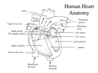
Heart Anatomy. Contents. Position. External morphology. Cardiac Chambers. Structure of the Heart. Conduction System. Vessels of the Heart. Pericardium. Position of the heart. Lies within the pericardium in middle mediastinum

E N D
Contents Position External morphology Cardiac Chambers Structure of the Heart Conduction System Vessels of the Heart Pericardium
Position of the heart • Lies within the pericardium in middle mediastinum • Behind the body of sternum and coastal cartilages 2 to 6 • In front of thoracic vertebrae 5 to 8 • A third of it lies to the right of median plan and 2/3 to the left
External morphology • one apex, • one base, • two surfaces • three borders • three grooves
Atria Auricles
Anterior Interventricular Sulcus Posterior Interventricular Sulcus
Left Ventricle Right Ventricle
Cardiac chambers • right atrium • right ventricle • left atrium • left ventricle
Pectinate Muscle
Right ventricle tricuspid valve
Papillary muscles Trabeculae carneae
Left ventricle mitral valve
Left ventricle inflow and outflow tracts (divided by the ant.cusp of the bicuspid valves) ※the papillary muscles(2 groups)
Heart Valves Bicuspid (mitral) valve Aortic valve Pulmonary valve Tricuspid valve
Semilunar valve Ventriclesystole Ventricle diastole
Tricuspid valve & mitral valve mitral valve tricuspid valve
tricuspid bicuspid
想一想… Ventricle diastole or systole ?
想一想… Ventricle diastole or systole ?
Structure of the heart the walls of the heart 3 layers — endocardium *continue with the lining of the large blood vessels — myocardium *2 kinds: the ordinary cardiac muscles the specially m. — epicardium • Septum interatrial septum ----Oval fossa interventricular septum ----Membranous part
Structure the walls of the heart 3 layers —endocardium *continue with the lining of the large blood vessels —myocardium *2 kinds: the ordinary cardiac muscles the specially m. — epicardium • Septum interatrial septum ----Oval fossa interventricular septum ----Membranous part
Atria Septum Ventricles
Left ventricle
Conduction System It is consists of the special cardiac muscles 5 parts: Sinoatrial node Atrioventricular node, Atrio-ventricular bundle Left and right branches Purkinje fibers.
— The sinoatrial node (SAN) *It is lies the junction between the right auricle and the sup.vena cava — The atrioventricular node (AVN) *It is lies in the lower portion of the interatrial septum just above the orifice of the coronary sinus — Purkinje fibers
Vessels of Heart • The arteries the left coronary a. the right coronary a. • The veins of the heart the coronary sinus the ant. cardiac v. the smallest v.
right coronary a. left coronary a.
The arteries The left coronary a. • arises from the left aortic sinus • 2 branches: ant.interventricular br. and circumflex br. • To supply the left atrium, left ventricle, the anterior surface of the right ventricle,anterior 2/3 of the interventricular septum,sometimes, supply the SAN and AVN.
The arteries The right coronary a. • arise from the right aortic sinus and runs along the right portion of the coronary groove • 2 branches: post interventricular branch post branch of the left venticle • To supply right atrium, right ventriclepost. 1/3 of the interventricular septum, the diaphragmatic surface of left ventricle,the SAN and AVN.
coronary artery bypass graft 冠状动脉搭桥术
The arteries The coronary sinusIt lies in the post.portion of the coronary sulcus between the left atrium and left ventricle.It opens into the right atrium.It receives:the great cardiac v.,the middle cardiac v. and small cardiac v.The ant. cardiac v.The smallest v.