Chapter 20
Chapter 20. Cardiovascular System The Heart. The Heart. The Heart is two pumps. Pulmonary circulation Carries blood to lungs Returns to the left side of heart Systemic circulation Delivers oxygen and nutrients to the body Returns to the right side of the heart. Functions of the Heart.

Chapter 20
E N D
Presentation Transcript
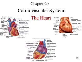
Chapter 20 Cardiovascular System The Heart
The Heart • The Heart is two pumps. • Pulmonary circulation • Carries blood to lungs • Returns to the left side of heart • Systemic circulation • Delivers oxygen and nutrients to the body • Returns to the right side of the heart
Functions of the Heart • Generating blood pressure • Routing blood • Heart separates pulmonary and systemic circulations • Ensures oxygenation of blood flowing to tissues. • Ensuring one-way blood flow • Heart valves ensure one-way flow • Regulating blood supply • Changes in contraction rate and force match blood delivery to changing metabolic needs
Size, Shape, Location of the Heart • Size of a closed fist • Shape • Apex: rounded tip of heart • Base: Flat part at opposite of end of cone • Located in thoracic cavity within the mediastinum
Heart Wall • Three layers of tissue • Epicardium: This serous membrane of smooth outer surface of heart • Myocardium: Middle layer composed of cardiac muscle cell and responsibility for heart contracting • Endocardium: Smooth inner surface of heart chambers
External Anatomy • Four chambers • 2 atria • 2 ventricles • Auricles • Major veins • Superior vena cava • Inferior vena cava • Pulmonary veins • Major arteries • Aorta • Pulmonary trunk
Heart Valves • Atrioventricular • Tricuspid • Bicuspid or mitral • Semilunar • Aortic • Pulmonary • Prevent blood from flowing back
Heart Skeleton • Consists of plate of fibrous connective tissue between atria and ventricles • Fibrous rings around valves to support • Serves as electrical insulation between atria and ventricles • Provides site for muscle attachment
Cardiac Muscle • Elongated, branching cells containing 1-2 centrally located nuclei • Contains actin and myosin myofilaments • Intercalated disks: Specialized cell-cell contacts • Desmosomes hold cells together and gap junctions allow action potentials • Electrically, cardiac muscle behaves as single unit
Electrical Properties • Resting membrane potential (RMP) present • Depends on low permiability to Na+ and Ca++ and high permiability to K+. • Action potential • Depolarization (Fast voltage gated Na+ channels) • Early partial repolarization (Voltage gated Na+ channels close and a small number of K+ channels open.) • Plateau phase - Prolonged period of slow repolarization (Slow Ca++ channels are open) • Rapid final repolarization phase (Voltage gated Ca++ channels close and more K+ channels open)
SA Node Action Potential Permiability changes in the pacemaker cells (Autorhythmicity): • Prepotential • Small number of Na+ channels open • Voltage-gated K+ channels are closing • Voltage-gated Ca++ channels begin to open • Depolarization Phase • Voltage-gated Ca++ channels are open • Voltage-gated K+ channels are closed. • Repolarization phase • Voltage-gated Ca++ channels close. • Voltage-gated K+ channels open. Note: Ca++ channel blockers like verapamil are used to treat tachycardia and arrhythmias.
Refractory Period • Absolute: Cardiac muscle cell completely insensitive to further stimulation • Relative: Cell exhibits reduced sensitivity to additional stimulation • Long refractory period prevents tetanic contractions
Electrocardiogram • Action potentials through myocardium during cardiac cycle produces electric currents than can be measured • Pattern • P wave • Atria depolarization • QRS complex • Ventricle depolarization • Atria repolarization • T wave: • Ventricle repolarization
Cardiac Arrhythmias • Tachycardia: Heart rate in excess of 100bpm • Bradycardia: Heart rate less than 60 bpm • Sinus arrhythmia: Heart rate varies 5% during respiratory cycle and up to 30% during deep respiration • Premature atrial contractions: Occasional shortened intervals between one contraction and succeeding, frequently occurs in healthy people
Cardiac Cycle • Heart is two pumps that work together, right and left half • Repetitive contraction (systole) and relaxation (diastole) of heart chambers • Blood moves through circulatory system from areas of higher to lower pressure. • Contraction of heart produces the pressure
Heart Sounds • First heart sound or “lubb” • Atrioventricular valves and surrounding fluid vibrations as valves close at beginning of ventricular systole • Second heart sound or “dupp” • Results from closure of aortic and pulmonary semilunar valves at beginning of ventricular diastole, lasts longer • Third heart sound (occasional) • Caused by turbulent blood flow into ventricles and detected near end of first one-third of diastole • Clinical considerations: • Murmurs: abnormal heart sounds • Incompetent valve - leaks excessively (gurguling,swishing) • Stenosis: abnormally small valve openings (rushing sound before valve closes. • Causes: genetic or due to rhematic fever scaring or myocardial infarction affecting the papillary muscles.
Mean Arterial Blood Pressure • Stroke volume • Volume of blood pumped during each cardiac cycle • at rest ~70 ml • can increase to ~200 ml during exercise • Heart Rate • ~72 BPM and can increase to over 120 BPM during exercise. • Cardiac Output =stroke volume X heart rate • Resting: 72 beats/min X 70 ml/beat = 5040 ml / min • Exercise: 120 beats/min X 200 ml/beat = 24,000 ml/min. • Cardiac reserve • max cardiac output - resting cardiac output • = 24,000-5040ml /min = 18960 ml • C.O. is the major factor in determining blood pressure. • Blood pressure • Blood pressure reflects pressure changes in the aorta not the ventricle • Normal BP 120 systolic / 80 diastolic
Regulation of the Heart • Intrinsic regulation: Results from normal functional characteristics, not on neural or hormonal regulation • Starling’s law of the heart • Stretching of the SA node. • Extrinsic regulation: Involves neural and hormonal control • Parasympathetic stimulation • Supplied by vagus nerve, decreases heart rate, acetylcholine secreted • Sympathetic stimulation • Supplied by cardiac nerves, increases heart rate and force of contraction, epinephrine and norepinephrine released
Heart Homeostasis • Effect of blood pressure • Baroreceptors monitor blood pressure • Effect of pH, carbon dioxide, oxygen • Chemoreceptors monitor • Effect of extracellular ion concentration • Increase or decrease in extracellular K+ decreases heart rate • Effect of body temperature • Heart rate increases when body temperature increases, heart rate decreases when body temperature decreases
Effects of Aging on the Heart • Gradual changes in heart function, minor under resting condition, more significant during exercise • Hypertrophy of left ventricle • Maximum heart rate decreases • Increased tendency for valves to function abnormally and arrhythmias to occur • Increased oxygen consumption required to pump same amount of blood