Download
1 / 15
150 likes | 242 Views
Explore the anatomy of muscles in the posterior neck region, innervation details, and palpation techniques to ensure safe assessment and care. Learn about the functions, structures, and potential discomfort areas of key muscles like Trapezius, Splenius, and Levator Scapulae. Take a closer look at the superficial and deep muscles, their shapes, and functions, with essential tips for palpation. Carefully understand the relation between muscle actions, nerve innervation, and potential trigger points for better muscle health management.

E N D
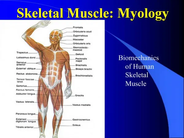
Myology Muscles of the Posterior Neck
Muscles of Neck Overview • Miscellaneous • Care must be taken when palpating anterior neck musculature. • The carotid sinus of the common carotid, if compressed, can cause a neurological reflex that decrease heart rate. This is especially important with weak and/or elderly clients. • The trachea, laryngeal cartilages, and thyroid gland are delicate. • Transverse processes (TP's) are bony and may cause discomfort if soft tissue is compressed into them.
Muscles of Neck Overview • Innervation • Trapezius and sternocleidomastoid (SCM) are both innervated by spinal accessory nerve (CN XI). • Suboccipitals are innervated by suboccipital nerve. • Scalenes, prevertebrals, and splenius capitis/cervicis are innervated by cervical spinal nerves. • Platysma is involved with facial expression and therefore innervated by the facial nerve (CN VII). • Infrahyoids innervated by cervical nerves. • Suprahyoids innervated by cranial nerves.
Muscles of the Posterior Neck – Superficial (4) Trapezius: Name tells us the left and right trap together have a trapezoid shape (diamond shape). Entire trap is superficial in neck and also in the back Directly deep to trap in neck are semispinalis capitis, splenius capitis & levator scapulae. Directly deep to trapezius in trunk are rhomboids and superior part of latissimus dorsi. Directly anterior to the anterior border of trap are levator scapulae and scalenes. Considered to have three functional parts: upper (which elevated the scapulae), middle (which adducts the scapulae), and lower (which depress the scapulae).
Muscles of the Posterior Neck – Superficial (4) • Splenius Capitis: • Shaped like a bandage (narrow rectangle) and attaches onto the head • Left & right splenius capitis muscles bilaterally form a “V” shape; sometimes known as the “golf tee” muscles • Splenius Cervicis: • Left & right splenius capitis muscles bilaterally form a “V” shape • Levator Scapulae: • At midpoint, there is a twist in the fibers that creates an increased density which is often mistaken for a trigger point (when present a levator scapulae trigger point is usually located just superior to the angle of the scapula). • Levator scapulae can also cause downward rotation of glenohumeral joint.
Trapezius O: EOP, Superior Nuchal Line, Nuchal ligament, and the SP of C7 through T12 I: Upper Traps: Lateral 1/3 of clavicle & Acromion Middle Traps: Spine of scapula and acromion Lower Traps: Root of the Spine of the scapula A: Upper Traps: Elevates, upwardly rotates, and retracts the scapula Middle Traps: Retracts the scapula Lower Traps: Depresses, upwardly rotates, and retracts the scapula **Reversed muscle action: Bilaterally allows for extension of the neck. Unilaterally laterally flexes the neck to the same side and rotates to the opposite side. N: CN XI (Spinal accessory nerve) and posterior rami of C3 and C4 Palpation: Page 111
Splenius Capitis O: Nuchal ligament and the SP’s of C7-T4 I: Mastoid process and the occiput A: Bilateral contraction: Extension of the neck Unilateral contraction: Lateral flexion and Ipsilateral rotation of the neck N: posterior rami of the cervical spinal nerves Palpation: Page 115
Splenius Cervicis • O: SP’s of T4 – T6 • I: TP’s of C1 – C3 • A: Bilateral contraction: Extension of the neck • Unilateral contraction: Lateral • flexion and Ipsilateral rotation of the neck • N: posterior rami of the cervical spinal nerves Palpation: Page 118
Levator Scapulae O: TP’s of C1 – C4 I: Medial border of the scapula, from the superior angle to the root of the spine of the scapula A: Elevates retracts, and downwardly rotates scapula. **Reversed muscle action: Bilaterally allows for extension of the neck. Unilaterally laterally flexes the neck to the same side and rotates the to the same side. N: Dorsal Scapular nerve Palpation: Page 118
Muscles of the Posterior Neck – Deep (Suboccipitals) (4) • Suboccipitals are found deep to trap, SCM, splenius capitis, and semispinalis capitis. • Suboccipitals are more important as postural muscles, providing fine control of head posture, than movers. • Rectus Capitis Posterior Major: • “rectus” means straight. Both rectus muscles run straight up to inferior nuchal line of occiput. • “capitis” refers to head • “posterior” means toward back • “major” means larger • Rectus Capitis Posterior Minor: • “minor” means smaller • Obliquus Capitis Inferior: • “obliquus” means slanted. Both obliquus muscles run in a slanted fashion. • Obliquus Capitis Superior
Rectus Capitis Posterior Major O: SP of the Axis (C2) I: Occiput (lateral aspect) A: Bilateral contraction: Extension of Head Unilateral contraction: Lateral flexion and Ipsilateral rotation of neck N: Suboccipital nerve Palpation: Page 127
Rectus Capitis Posterior Minor O: Posterior tubercle of the Atlas (C1) I: Occiput A: Bilateral contraction will cause Extension of Head N: Suboccipital nerve Palpation: Page 130
Obliquus Capitis Inferior O: SP of the Axis (C2) I: TP of the Atlas (C1) A: Ipsilateral Rotation of Atlas N: Suboccipital nerve Palpation: Page 132
Obliquus Capitis Superior O: TP of the Atlas (C1) I: Occiput (between the superior and inferior nuchal lines) A: Bilateral Contraction: Extension of Head Unilateral Contraction: Lateral flexion of the head. N: Suboccipital nerve Palpation: Page 135
Suboccipital Triangle • Formed by RCPM, OCS, & OCI, the suboccipital triangle is covered by a layer of dense fibro-fatty tissue, situated beneath the Semispinalis capitis. • The floor is formed by the posterior occipito-atlantal membrane, and the posterior arch of the atlas. • In the deep groove on the upper surface of the posterior arch of the atlas are the vertebral artery and the first cervical or suboccipital nerve.