Download
1 / 39
390 likes | 476 Views
Explore the different types of muscular tissues and their structures, functions, origins, and actions, along with the nomenclature and accessory structures of muscles. Discover the morphology and gross structures of skeletal muscles.

E N D
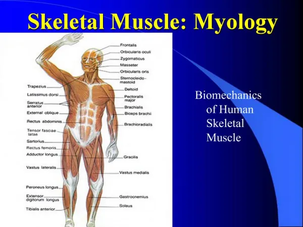
Section 1 The general descriptionA. Three variations of muscular tissue1. According to the gross structure1)skeletal muscle 2) smooth muscle 3) cardiac muscle2. According to the microscopic structure1)striped(striated) muscle (skeletal muscle and cardiac muscle) 2)unstriated muscle (smooth muscle)3. According to the function1)voluntary muscle (skeletal muscle) 2) involuntary muscle (smooth and cardiac muscles)
B. The morphology of skeletal muscles1.4 kinds of skeletal muscleslong, short, broad, orbicular muscles.2.The direction of muscular fibersbipennate muscles unipennate muscles multipennate muscles3. The tendinous numberdigastric m., biceps m., triceps m., quadriceps m..
bipennate muscles unipennate muscles multipennate muscles digastric m., biceps m., triceps m., quadriceps m..
bipennate muscles unipennate muscles multipennate muscles
C.The gross structures of a skeletal muscle: 1. Belly and tendon : aponeurosis, tendinous intersection intermediate tendon digastric m. triceps m. biceps m. quadriceps m.
D.The origin, insertion of skeletal muscle: 1.The fixation of muscles:most muscles are attached to bones, cartilages, ligaments, fasciae or combination of them, some are attached to organs, skin or mucous membrane. 2. Origin and insertion: a) in general, when a muscle contracts or shortens, fixed attachment is origin, the moveable one is insertion, so in limbs, the distal parts of muscles usually are insertion. b) sometimes the flexion and movement parts of the muscle may be exchanged each other.
E.The action of the muscles: 1. Prime movers or agonists: contract actively 2. Antagonists: oppose the action of a prime mover 3. Synergist: cooperate in performing an action 4. Fixators
F.The nomenclature of muscles: The names of muscles indicated shape, location, actions or their combination of muscles.
The nomenclature of the muscleThe names of muscles indicated shape, location, actions or their combination of muscles.
G.The accessory structures of the muscles 1.The fascia: a) superficial fascia(fat, the trunks of subcutaneous vessels and nerves, the superficial lymph notes, the mammary gland and cutaneous muscles.) b) deep fascia (proper fascia) ---form some sheaths for each or group of muscles and for vessels and nervous. ---intermuscular septa separating the groups of muscles ---retinaculum for its underlying tendons.
G. The accessory structures of the muscles1. The fascia1)The superficial fascia --- immediately beneath the cutis, covering almost the entire body. --- to be composed of loose connective tissue containing fat in varying quantity. --- varies in thickness in different individuals and different parts of the body.--- contains the trunks of the subcutaneous vessels and nerves, the superficial lymph nodes the mammary gland and certain cutaneous muscles.
2)The deep fascia (proper fascia)--- forms some sheaths for each or group of muscles and for vessels and nerves. --- intermuscular septa separating the groups of muscles. --- retinaculum for its underlying tendons.
2.The synovial tendon sheath (or tendinous sheath): 2 layers a) fibrous layer (or fibrous sheath of tendon) b) synovial layer (or synovial sheath of tendon) --- parietal layer and visceral layer (synovial fluid) --- mesotendon (mesotendineum) carries blood vessels to the tendon.
3. The synovial bursa 4. The sesamoid bones
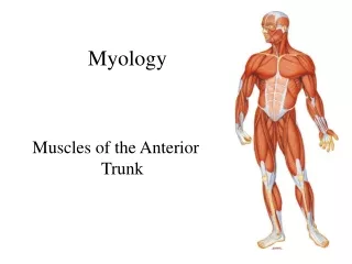
Section 2 The Muscles of Trunk. The muscles of backtrapezius, latissimus, levattor scapulae, rhomboid muscles erector spinae (sacrospinelis) thoracolumbar fascia. The muscles of thorax1) The extrinsic muscles pectoralis major, pectoralis minor, serratus anterior 2) The intrinsic muscles intercostales extrerni, intercostales interni . The diaphragm . The muscles of abdomen
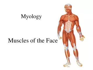
Section 3 The Muscles of Head and Neck. The muscles of head1. The facial muscles2. The masticatory muscles. The muscles of neck1. The superficial groupplytama, sternocleidomastoid 2. The hyoid muscle 1) The suprahyoid muscles digastric, mylohyoid, stylohyoid, geniohyoid2) The infrahyoid muscles sternohyoid, omohyoid; sternothyoid, thyrohyoid