Download
1 / 79
920 likes | 1.64k Views
Rheumatoid Arthritis. Introduction. Epidemiology/genetics Pathogenesis Clinical Features Laboratory Manifestations Diagnosis Management considerations and therapy. Rheumatoid arthritis. Chronic, systemic inflammatory disease Unknown etiology Persistent inflammatory synovitis

E N D
Introduction • Epidemiology/genetics • Pathogenesis • Clinical Features • Laboratory Manifestations • Diagnosis • Management considerations and therapy
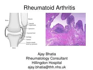
Rheumatoid arthritis • Chronic, systemic inflammatory disease • Unknown etiology • Persistent inflammatory synovitis • Synovial inflammation (pannus) cartilage destruction, bone erosion with subsequent deformity • Peripheral joints in symmetric fashion • Extra-articular manifestations also occur
Epidemiology • 2.5 million Americans (~1%), 165 million worldwide • Females > males 3:1; all races • Peak age onset: 4th-5th decade • 80% develop between ages 35-50yrs • Prevalence increases with age
Genetics • Strongly associated with HLA-DR4 • DRB1*0401/0404 severe and erosive disease • “Shared epitope” on 3rdhypervariable region of HLA-DRB1 • + HLA-DR4 seen in 20-30% of general population • Other factors involved for disease to develop
Genetics • T lymphocytes recognizing antigens in synovial tissue • T cells, macrophages + fibroblasts produce pro-inflammatory cytokines • Play a key role in synovitis and tissue destruction • Pro-inflammatory cytokines: TNF alpha, IL-1 and IL-6
Pathophysiological Role of Cytokines + Other Mediators and Inhibitors in RA Scott D and Kingsley G. N Engl J Med 2006;355:704-712.
Risk Factors For Aggressive RA • HLA-DR4, High titer RF and + CCP ab • Early radiographic erosions • Constitutional symptoms • Insidious onset • Early appearance of rheumatoid nodules
Disease onset • Insidious: Most common presentation • Small peripheral joints: MCPs, PIPs, wrists • Abrupt: • Acute polyarthritis Intense pain, swelling + limitation • Slow monoarticular: • Knees/shoulders progresses to small joints
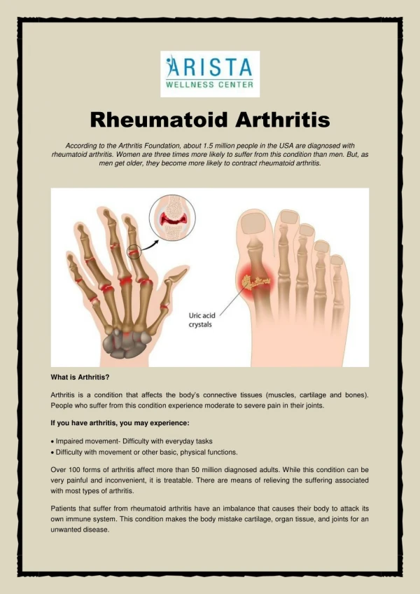
Overview: Joint Involvement TMJ: 20-30% C-spine: 40-50% Shoulder: 50-60% SC Joints : ? Elbow: 40-50% Wrist: 80-90% Hand: MCP 90-95% PIP 65-90% Hips: 40-50% Knees: 60-80% Ankles: 50-80% Foot : MTP 50-90% PIP 65-90% www.moviesyahoo.com
Clinical features • Symmetric inflammatory synovitis(palpable swelling) of small peripheral joints • Tenderness to palpation and ROM • Symptoms last > 6 weeks • Useful indicator of disease activity • Morning stiffness > 1˚ improves with activity
Clinical features • Symmetric polyarticularjoint involvement (>3 joints) • Small joint arthropathy:MCPs, PIPs, wrists, MTPs • Knees, ankles and shoulders • Typically spares: thoracolumbar spine + DIP joints • Entrapment syndromes also commonly occur • Carpal tunnel and tarsal tunnel
Boutonniere Deformity www.medscape.com www.medscape.com
Swan neck deformity www.medscape.com
Baker’s cyst • Swelling posterior knee • Ruptured popliteal cyst swelling of calf (pseudo-phlebitis) • Mimics DVT • “Crescent sign”
Axial Disease • Anterior alantoaxialsubluxation of C1-2 common • ≥ 3 mm separation between odontoid and atlas • Recurrent HA, tingling in UE, unexplained dizziness • Susceptible to trauma with endotracheal intubation • Must get pre-op X-rays of neck (lateral flexion/extension) • Symptomatic cervical myelopathy spinal fusion
Extra-articular manifestations • 40% of patients • Increased frequency: • +RF, +CCP ab, HLA-DR1 +DR4 • Environmental factors such as smoking • Life expectancy loss of 18 yrs • 5x mortality risk
Rheumatoid nodules • 20-40%of SPRA patients • Reflects level of RA disease activity • Develops on pressure areas • Risk factors: +RF, subchondral cysts, Methotrexate(MTX)
Rheumatoid nodules • Single/multiple nodules • Interfere with function/ulcerate • Regress with DMARDS • MTX may result in ↑nodulosis
Rheumatoid nodules • May involve internal organs • Sites of movement: • Pulmonary parenchyma/pleura • Pericardium/myocardium • Heart valves • Vocal cords
Caplan’s syndrome • Pulmonary nodulosis + pneumoconiosis • Exposure to inorganic dusts (coal, asbestos, silca) • Similar to simple rheumatoid nodules • Modified tissue response to inhaled dusts • May lead to progressive massive fibrosis (PMF)
Interstitial lung disease • Most common lung manifestation • ↑mesenchymal reactivity fibrosis • PE: fine, diffuse dry rales; low DLCO • CXR: • Reticular/reticulonodular pattern honeycombing
Interstitial lung disease • Wide spectrum of findings on lung biopsy • Histologic finding idiopathic interstitial pneumonia (IIP) • Tx: “ground-glass” on HRCT good response to tx • High dose steroids, imuran and cytoxan
Pleurisy/pleural disease • Inflamed pleura thicken, calcify + forming adhesions • Pleural fluid reveals: • Low glucose (< 30mg/dl) • High protein (>4 g/dl) and LDH • Low complement (CH50 ) • Cellular infiltrates (mononuclear) • Improves with treatment of RA www.uptodate.com
Hematologic involvement • Mild hypochromic normocytic anemia • Thrombocytosis • Lymphadenopathy • Felty’s syndrome • Large granular lymphocyte syndrome • “Pseudo-Felty Syndrome
Felty’s syndrome • Classic triad: RA, neutropenia, splenomegaly • Risk factors: RF+, nodular RA and +HLADR4 • Manifestations: • Non-healing leg ulcers • Infections • PMNs < 1000/mm3 • Common cause of death www. knol.google.com
Felty’s syndrome • Treatment: • DMARDs Methotrexate • Splenectomy • TNFi no studies in actual treatment of Felty’s • Steroids improve neutropenia but ↑ risk of infection
Large granular Lymphocyte syndrome • Variant of Felty’s • Peripheral blood or bone marrow LGL cells • Circulating LGLs, neutropenia, frequent infections, splenomegaly • 3-14% leukemia unlike Felty’s. No splenectomy!
Ocular Involvement • Keratoconjunctivitissicca • Episcleritis • Local or diffuse • Scleritis • Local or diffuse • Scleromalaciaperforans • Choroid and retinal nodules
Rheumatoid vasculitis • < 1% of RA pts • Risk factors: • High titer RF & long standing, severe disease (> 10yrs) • Male gender • Smoking • Prior DMARD use • Hypocomplemetemia • Circulating cryoglobins
Rheumatoid vasculitis • Clinical presentations: • Cutaneous ulcerations • Mononeuritis multiplex • Foot/wrist drop • Palpable purpura • Distal arteritis • Visceral arteritis: • Heart, lungs, bowel, spleen, kidneys
Course of RA • Intermittent (15-20%) • Long clinical remission (10%) • Progressive disease (65-70%)
Risk factors for increased morbidity/mortality Physical factors: • Extra-articular features • Erosions on x-ray • ↑RF + ↑ESR/CRP • Duration of disease • Disability at diagnosis • > 20 swollen joints Social factors: • Early age at diagnosis • ↓ socioeconomic status • Psychosocial stress • Low HAQ scores
Morbidity/Mortality in ra • Median life expectancy decreased by 3-18 yrs • Mortality rates higher with extra-articularmanifestions and women • ~50% stop working within 5-10ys of diagnosis • ~80% disabled to some degree after 20 yrs
Morbidity/Mortality in ra • Infection: 70% more likely to have infection • Don’t forget septic arthritis in RA patients arthocentesis • NH lymphoma: 2-5 fold increased risk • CAD: 3x the risk of sudden death/MI • Cerebrovascular diseases: • 70% more likely to have a stroke
Laboratory manifestations • No lab test is specific for RA • RF + • Anti-CCP + (anti-cyclic citrullinated peptide antibody) • Increased ESR/CRP • ANA + (25% of patients) • Anemia +thrombocytosis
Rheumatoid factor • Usually IgMAb recognizes Fc portion of IgG molecule • 70% RF+ at onset, 85% overall in first 2 years • High titer severe disease, extra-articular manifestations, increased mortality • Normal 1-4%, 10-25% + over age 70
Mnemonic for +RF • CHronic: • CH: Chronic diseases liver/pulmonary/sarcoidosis • R: Rheumatoid arthritis • O: Other CTDS (SLE, SS, MCTD) • N:Neoplasms (XRT, chemotherapy) • I:Infections (SBE, HIV, Hepatitis B+C, TB, Parvovirus B19) • C:Cryoglobinemia
Anti-CCP • Autoantibody directed at cyclic citrullinated peptide • Sensitivity 65-70%, specificity (95%) • RF and CCP ab combination specificity 99.5% • Detectable in early RA • May antedate onset of inflammatory disease • Predictor of aggressive + erosive disease
1987 ACR classification criteria for RA Must have 4 of 7 criteria: • Morning stiffness at least 1 ˚ • Swelling in 3 or more joints • Swelling of MCP, PIP or wrist joints • Symmetric joint swelling • Radiographic erosions or periarticular osteopenia • SQ rheumatoid nodules • Positive RF At least 6 weeks