Download
1 / 32
320 likes | 399 Views
Learn about diagnosing fractures through clinical features and imaging techniques, and understand the healing process for natural bone repair. Explore the various types of fractures and appropriate approaches to handling them.

E N D
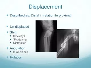
Displacement • Described as: Distal in relation to proximal • Un-displaced • Shift • Sideways • Shortening • Distraction • Angulation • In all planes • Rotation
Fracture Diagnosis • Clinical features • Imaging: Radiology (x-Ray)
Clinical Features • History of Trauma • Symptoms and signs: • Pain • Swelling • Deformity • Bony tenderness • Abnormal movement • Crepitus • Loss of function
Approach - history • Details of injury • Mechanism, force, bleeding, consciousness, … • Details of facture • Deformity, pain, loss of function, .. • Other medical problems • Anti-tetanus status if open injuries • Careful: • Fractures are not always at the site of impact • Some fractures do not need severe force
Approach – clinical exam • General medical condition • should be evaluated to exclude • shock • brain injury • other problems • Vital signs • should be observed and followed up
Approach – clinical exam • Look: • Adequate exposure • General on patient • Local: • Swelling, deformity, bruises, color, … • Special attention is to be paid to wounds
Approach – clinical exam • Feel: • Tenderness, distal pulses, temperature and crepitus on movement • Sensory and motor deficits • Pulse distal to injury • Compartment syndrome • Move: • With care • make sure not to cause more pain or injury • Crepitus &abnormal movement indicates a fracture • Joints distal to the affected area
Approach – clinical exam • Examination of the viscera • Liver and spleen in rib fractures • Urinary bladder and urethra in pelvic fractures • Neurological examination in head and spinal injury
Investigations - Imaging • X-rays: • Low of 2s • Two views: AP and Lateral • Two joints: Above and Below • Two sides: Right and Left • Two occasions • Two Doctors ! • Special views: • Obliques, Tunnel view, skyline, functional flexion / extension • Arthrography: • Shows intra-articular structures • Functional in hip
Imaging • Plain x-ray:(law of twos) • Two views:AP and Lateral AP Lat AP Lat Apley’s System of Orthopedics & Fractures
Imaging • Plain x-ray:(law of twos) • Two views: AP and Lateral • Two joints: joint above and joint below Apley’s System of Orthopedics & Fractures
Imaging • Plain x-ray:(law of twos) • Two views: AP and Lateral • Two joints: joint above and joint below • Two limbs: for comparison • more in children to compare epiphysis Apley’s System of Orthopedics & Fractures
Imaging • Plain x-ray:(law of twos) • Two views: AP and Lateral • Two joints: joint above and joint below • Two limbs: for comparison • more in children to compare epiphysis • Two occasions • e.g. stress fractures • e.g. scaphoid fracture Apley’s System of Orthopedics & Fractures
Imaging • Plain x-ray:(law of twos) • Two views: AP and Lateral • Two joints: joint above and joint below • Two limbs: for comparison • more in children to compare epiphysis • Two occasions • e.g. stress fractures • e.g. scaphoid fracture • Two injuries • e.g. patellar fracture and hip injury • e.g. calcaneal fractures & spine injuries www.jumpintheair.com
Imaging • Plain x-ray:(law of twos) • Two views: AP and Lateral • Two joints: joint above and joint below • Two limbs: for comparison • more in children to compare epiphysis • Two occasions • e.g. stress fractures • e.g. scaphoid fracture • Two injuries • e.g. calcaneal fractures & spine injuries • .....and two Doctors! www.123rf.com/
Imaging • Plain x-ray: (law of twos) • Special views: • Calcaneal view • Shoulder dislocation: axial view • Scaphoid views • Acetabular fractures: 45o tilt views http://osuemed.wordpress.com/
Imaging • CT Scan: • In complex and ntra-articular fractures • In spine • In pelvic and acetabular fractures • In calcaneal fractures www.learningradiology.com
Imaging • MRI • Show associated injuries in spinal fractures • Associated soft tissue injuries – e.g. knee • Hidden fractures: • Subtrochanteric (ST) disruption • Stress (fatigue) fractures • Scaphoid fracture • Suspected avascular necrosis www.highperformancesports.blogspot.com www.bjj.boneandjoint.org.uk
Fracture healing • A broken bone heels because …..it is broken ! Alan Apley
Natural bone healing • Movement at the fracture site initiates a healing process—callus formation • Vascular and cellular response leads to tissue differentiation and mineralization resulting in restoration of mechanical integrity
Natural bone healing http://classes.midlandstech.edu/
Cascade of tissue differentiation Following a Fracture: • Hematoma • Granulation tissue • Connectivetissue • Fibrocartilage • Mineral deposition • Bone
bony bridging Fracture healing • Inflammation • Hematoma • Mesenchymal cells • Soft callus • Granualation tissue • Hard callus • Intramembranous bone formation • Enchondral ossification • Remodeling
Cellular and Vascular Reaction cells haematoma granulation tissue
Tissue Differentiation connective tissue granulation tissue Giemsa
Tissue Differentiation Cascade Masson-Goldner Cartilage formation Mineral deposition Bone
Fracture Healing • Conditions necessary for bone healing: • Good blood supply • Controlled motion • No infection
Fracture Healing • Unfavorable factors • Impairment of blood supply • Infection • Excessive movement • Presence of tumor • Interposition of soft tissue • Any form of Nicotine (smoking) • Bad nutrition
Average healing time • Children: • Upper limb: 3-4 weeks • Lower limb: 2X upper limb (6-8 weeks) • Adults: • Upper limb: 2X children (6-8 weeks) • Lower limb: 2X upper limb (12-16 weeks)
Fracture Treatment • Aim of fracture treatment • aid healing, • in normal position, • avoiding complications
Fracture treatment • Treat the patient, not only the fracture • Reduce the fracture • Immobilize the fracture • Prevents displacement • Alleviates pain • Promotes soft tissue healing • Mobilize the patient • Avoid complications