EYELID DISORDERS
500 likes | 1.01k Views
EYELID DISORDERS. Instructor : Dr. Arkam DONE BY : Sofian bni awwad. Anatomy :. The eyelid is consist of four layers: 1- An anterior layer of skin and subcutaneous tissue.

EYELID DISORDERS
E N D
Presentation Transcript
EYELID DISORDERS Instructor : Dr. Arkam DONE BY :Sofianbniawwad
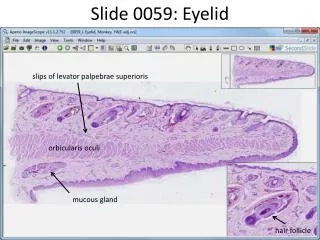
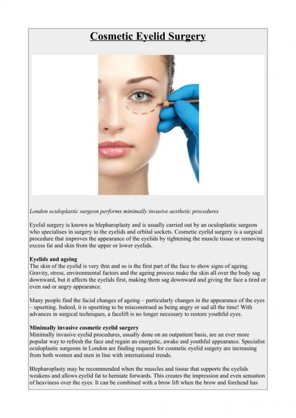
Anatomy : • The eyelid is consist of four layers: 1- An anterior layer of skin and subcutaneous tissue. 2- Muscular layer that comprises the orbicularisoculi muscle, which is responsible for the closing of the lids. 3- Tarsal plate which is a tough collagenous layer that houses meibomian gland. 4- Tarsal (palpebral )conjunctiva. • The orbital septum represents the anatomic boundary between the lid tissue and the orbital tissue. • Innervations: majorly by ophthalmic and maxillary branch of trigeminal nerve. • Blood supply: majorly by ophthalmic and lacrimal artery.
FUNCTION • It offers mechanical protection to anterior globe • Spread the tear film over the conjunctiva and cornea with each blink. • Contain the meibomian oil gland which provide the lipid component of the tear film. • Prevent drying of the eyes. • Contain the puncta through which the tears flow into the lacrimal drainage system.
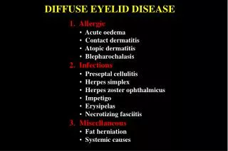
ptosis • This is an abnormally low position of the upper eyelid. PATHOGENESIS It may be caused by: Mechanical factors: • (a) Large lid lesions pulling down the lid. • (b) Lid oedema. • (c) Tethering of the lid by conjunctival scarring. • (d) Structural abnormalities including a disinsertion of the aponeurosis of the levator muscle, usually in elderly patients.
2.Neurological factors: • (a)Third nerve palsy • (b)Horner’s syndrome, due to a sympathetic nerve lesion • (c)Marcus–Gunn jaw-winking syndrome. 3.Myogenic factors: • (a)Myasthenia gravis • (b)Some forms of muscular dystrophy. • (c)Chronic external ophthalmoplegia.
SYMPTOMS Patients present because: • they object to the cosmetic effect; • vision may be impaired; • there are symptoms and signs associated with the underlying cause (e.g. asymmetric pupils in Horner’s syndrome, diplopia and reduced eye movements in a third nerve palsy).
Signs : • There is a reduction in size of the interpalpebral aperture. • The upper lid margin, which usually overlaps the upper limbus by 1–2imm, may be partially covering the pupil. • The function of the levator muscle can be tested by measuring the maximum travel of the upper lid from upgaze to downgaze (normally 15–18imm). Pressure on the brow (frontalis muscle) during this test will prevent its contribution to lid elevation. • If myasthenia is suspected the ptosis should be observed during repeated lid movement. Increasing ptosis after repeated elevation and depression of the lid is suggestive of myasthenia
MANAGMENT • It is important to exclude an underlying cause whose treatment could resolve the problem (e.g. myasthenia gravis). Ptosis otherwise requires surgical correction • In very young children this is usually deferred but may be speed up if pupil cover threatens to induce amblyopia.
Marcus Gunn Jaw-Winking syndrome - Also called Trigemino-oculomotorSynkineses • Autosomal dominant • In this congenital ptosis there is miswiring of the nerve supply to the pterygoid muscle of the jaw and the levator of the eye so that the eyelid moves in conjugation with movements of the jaw. Treatment • Treatment is usually unnecessary but in severe cases, surgery with a bilateral levator excision and frontalis brow suspension may be used.
dermatochalasis • excessive and lax eyelid skin and muscle is known as dermatochalasis. Gravity, loss of elastic tissue in the skin, and weakening of the connective tissues of the eyelid frequently contribute to this lax and redundant eyelid tissue. These findings are more common in the upper eyelids but can be seen in the lower eyelids as well. • The patients who complain of dermatochalasis frequently complain of visual difficulties • Causes: • The most common cause of dermatochalasis is the normal aging phenomenon • Patients with severe periorbital edema may develop dermatochalasis • Trauma can be associated with dermatochalasis • Chronic dermatitis • Thyroid eye disease • Chronic renal insufficiency • Amyloidosis • Genetics may play a role in some patients who develop dermatochalasis • Treatment: • Blepharoplasty is the procedure of choice for upper and/or lower eyelid dermatochalasis
Entropion • It is an inturning, usually of the lower lid towards the globe. - Patients present with irritation caused by eyelashes rubbing on the cornea. - more common in elderly, because orbcularis muscle become spasm. • it may also caused by Conjuctival scarring distorting the lid (cicatrical entropion) • Treatment: • Short term :include the application of lubricants to the eye or taping of the eyelid. • Permenant :surgery
Ectropion • Eversion of the lid away from the globe. • Causes:- -age related orbicularis muscle laxity. -facial nerve palsy. -scarring of periorbital skin. - initial complaint of watery eye, because the mal position of the lids everts the puncta and prevents drainge of the tears leading to epiphora(overflow of the tears over the cheeks ) -it also exposes the conjuctivaleading to irratableeye. • treatment: surgical
Blepharitis • Inflammation of the eyelid margins. • It is a chronic disease. • Symptoms: • tired, itchy, sore eye, worse in the morning. • Crusting of the lid margin. • Classified into: anterior and posterior . • Both forms are strongly associated with seborrhoeic dermatitis, atopic eczema and acne rosacea.
Anterior Blepharitis • Is when the inflammation is located in the outside surface the lid margin, specifically in lash line. • Signs are: -Redness and scaling of the lid margin. -Debris in the form of a collarette around the eyelashes. -Reduction in the number of eyelashes. -Some lash bases may ulcerated- sign of staphylococcal infection. • In severe diseases the cornea is affected (blepharokeratitis) • Small infiltrate ulcers may form in the peripheral cornea (marginal keratitis)due to immune complex response to staphlococcalexotoxins .
Posterior blepharitis • Have another name which is meibomian gland dysfunction. • Signs are: - Obstruction and plugging of the meibomian orifices. - Thickened , cloudy, expressed meibomian secretion. - Injection of the lid margin and conjuctiva. - Tear film abnormalities and punctuate keratitis.
Treatment Anterior blepharitis: • Cleaning with a cotton bud wetted with bicarbonate or diluted baby shampoo to remove squamous debris from lash line . • Topical steroid: used infrequently. • Topical (fusidic acid) +- systemic antibiotic in staphylococcal lid disease . Posterior blepharitis: • Hot compressors and lid massage. • Oral tetracycline. • Artificial tears to prevent dryness
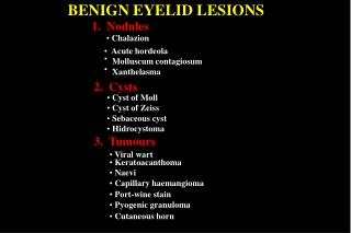
Chalazion • -It is a granuloma within the tarsal plate caused by obstructed meibomian gland. • -Painless. • -Symptoms are unsightly lid swelling which resolve within six months if the lesion persist we remove it surgically ,
Internal hordeolum • an abscess in meibomian gland. • -Painful. • -May respond to topical antibiotics but incision maybe necessary. ,
External Hordeolum (Stye) , . • - It is an abscess in eyelash follicle. • painful • -Most cases are self limiting . • -Treatment requires the removal of the associated eyelash and application of hot compresses.
Molluscum Contagiosum , , • -Is a viral infection of the skin or the mucous membranes, caused by pox virus. • -Can be presented with umbilicated lesion found on the lid margin. • -Cause irritation, redness, follicular conjuctivitis(small elevation of lymphoid tissue found on tarsal conjunctiva) • -Treatment requires excision of the lid lesion.
Xanthelasma • - Lipid containing bilateral lesions. • - Usually associated with hyperlipidemia . • - Removed for cosmetic reasons.
THANX FOR LISTENING DONE BY:Sofianbniawwad :”)