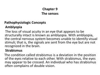
Amblyopia
E N D
Presentation Transcript
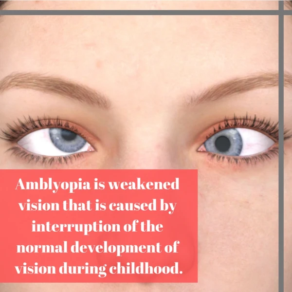
Unilateral or less commonly, bilateral reduction of best corrected visual acuity that can not be attributed directly to the effect of any structural abnormality of the eye or the posterior visual pathway.
Resulting from one of following: Strabismus - DEVIATION Anisometropia or high bilateral refractive error (Isoametropia) - DEFOCUS Visual deprivation - DEPRIVATION
Prevalence: 2%-4% Commonly unilateral Nearly all amblyopic visual loss is preventable or reversible with timely detection and appropriate intervention. Children with amblyopia or at risk for amblyopia should be identified at a young age when the prognosis for successful treatment is best. Role of screening is important
Amblyopia is primarily a defect of central vision. There is a critical period for sensitivity in developing amblyopia. The time necessary for amblyopia to occur during critical period is shorter for stimulus deprivation than for strabismus or anisometropia.
Neurophysiology: Cells of the primary visual cortex can completely lose their innate ability or show significant functional deficiencies Abnormalities also occur in neurons in the lateral geniculate body Evidence concerning involvement at the retinal level remains inconclusive
Classification: Strabismus Amblyopia :Deviation Anisometropia Amblyopia : Defocuss Amblyopia Due to bilateral high refractive error (isometropic) :Defocuss Deprivation Amblyopia :Deviation
Strabismus Amblyopia The most common form of amblyopia Strabismic amblyopia is thought to result from competitive or inhibitory interaction between neurons carrying the nonfusible inputs from the two eyes. Which leads to domination of cortical vision centers by the fixating eye and chronically reduced responsiveness to the nonfixating eye input.
Anisometropia Amblyopia Second in frequency It develops when unequal refractive error in the two eyes causes the image on the one retina to be chronically defocused. This condition is thought to result: Partly from the direct effect of image blur in the development of visual acuity. Partly from intraocular competition or inhibition
Mild hyperopic or astigmatic anisometropia (1.5D) mild amblyopia Mild myopia anisometropia (less than -2.5D) usually doesn't cause amblyopia unilateral high myopia (-6D) sever amblyopia visual loss.
Amblyopia Due to bilateral high refractive error (isometropia) isometropic amblyopia result from large, approximately equal, uncorrected refractive error in both eyes of a young child. Hyperopia exceeding 5D & myopia excess of 10 D risk bilateral amblyopia
Meridonial amblyopia: Uncorrected bilateral astigmatism in early childhood may result in loss of resolving ability limited to chronically blurred meridians.
Deprivation Amblyopia It is usually caused by congenital or early acquired media opacity. This form of amblyopia is the least common but most damaging and difficult to treat. In bilateral cases acuity can be 20/200 or worse.
In children younger than 6 years, dense congenital cataract that occupy the central 3 mm. or more of the lens must be considered capable of causing sever amblyopia. Similar lens opacities acquired after 6 years are generally less harmful.
Small polar cataracts & lamellar cataracts may cause mild to moderate amblyopia or may have no effect on visual development. Occlusion amblyopia is a form of deprivation caused by excessive therapeutic patching.
Diagnosis Characteristics of vision alone cannot be used to reliably differentiate amblyopia from other form of visual loss. The crowding phenomenon is typical for amblyopia but not uniformly demonstrable. Afferent pupillary defect are Characteristic of optic nerve disease but occasiinally appear to be present with amblyopia
Multiple assessment using a variety of tests or performed on different occasions are sometime required to make a final judgment concerning the presence and severity of amblyopia.
Binocular fixation pattern: It is a test for estimating the relative level of vision in the two eyes for children with strabismus who are under the age of about 3. This test is quite sensitive for detecting amblyopia but results can be falsely positive. Showing a strong preference when vision is equal or nearly equal in the two eyes, particularly with small angle strabismic deviations.
The modified Snellen technique directly measures acuity in children 3-6 years old. Often, however, only isolated letters can be used, which may lead to under estimated amblyopia visual loss. Croding bar may help alleviate this problem.
Crowding bar, or contour interaction bars, allow the examinator to test the crowing phenomenon with isolated optotype. Bar surrounding the optotype mimic the full of optotype to the amblyopia child. O E
Treatment Treatment of amblyopiainvolves the following steps: Eliminating (if possible) any obstacle to vision such as a cataract Correcting refractive error Forcing use of the poorer eye by limiting use of the better eye.
Cataract removal Cataracts capable of producing amblyopia require surgery without unnecessary delay. Removal of significant congenital lens opacities during the first 2-3 months of life is necessary for optimal recovery of vision. In symmetrical bilateral cases, the interval between operations on the first and second eyes should be no more than 1 week. Acutely developing severe traumatic cataracts in children younger than 6 years should be removed within a few weeks of injury, if possible.
Refractive correction In generally, optical prescription for amblyopic eyes should correct the full refractive error as determined with cyclopagic.
Occlusion and optical degradation Full time occlusion of the sound eye: Defined as occlusion for all or all but one waking hour. It is the most powerful means of treating of amblyopia by enforced use of the defective eye. The patch can either be left in place at night or removed at bedtime. Spectacle-mounted occluser or special opaque contact lenses can be used as an alternative to full-time patching if skin irritation or poor adhesion proves to be a significant problem
Full time patching should generally be used only when constant strabismus eliminates any possibility of useful binocular vision because full time patching runs a small risk of perturbing binocularity.
Part-time occlusion: Defined as occlusion for 1-6 hours per day. The children undergoing part time occlusion should be kept as visually active as possible when the patch is in place. Compliance with occlusion therapy for amblyopia declines with increasing age.
Penalization: A cycloplegic agent (usually atropine 1% or homatropine ) once daily to the better eye This form of treatment has recently been demonstrated to be as effective as patching for mild to moderate amblyopia.
E.B.M. Evidence Based Medicine Prospective, randomised • PEDIG, MOTAS & COCHRANE Eminence Based Medicine • Hopkins: weekend atropine • Scott [Iowa]: only full time
PEDIG P • E • Diatric ophthalmology • Investigator • Group North American Community based Ophthalmology and optometry
MOTAS • Monitored • Occlusion • Treatment of • Amblyopia • Study England Alistair Fielder
PEDIG:Amblyopia 6/30 - 6/120 6 h/d vs. all [or all -1] waking hours • Ages 3-7 • Can do reliable HOTV • 1h/d near activity 4mo: 4+ line improvement both groups Age / severity of amblyopia NOT relevant to outcome!
PEDIG:Amblyopia 6/12- 6/24 2h vs. 6h/d opaque occluder • Ages 3-7 • Can do reliable HOTV • 1h/d near activity 4mo: same 2.4 line improvement Age / severity of amblyopia NOT relevant to outcome!
PEDIG:Amblyopia 6/12 - 6/24 Daily atropine vs. patch 6h/d • 6mo: no difference • Patch: faster response • 2y: amblyopic eye 1.8 lines worse in each group • Improvement @ 2y: 3.6 vs. 3.7 lines
PEDIG:Recurrence of amblyopia after stopping treatment ≥ 3 lines acuity improvement • 25%: ≥ 2 lines loss @ 12mo • 42% after stopping 6h/d • 14% if 6h/d tapered to 2h/d before stopping
MOTAS investigators:Recurrence of amblyopia after stopping treatment Factors affecting the stability of visual function following cessation of occlusion therapy for amblyopia. Graefe 6/2007 Tacagni DJ, …Fielder AR
MOTAS investigators:Recurrence of amblyopia after stopping treatment 1 y follow-up from treatment cessation: children with "mixed" amblyopia (both anisometropia and strabismus) had significantly (p=0.03) greater deterioration in VA (0.11+/-0.11 log units) than children with only anisometropia (0.02+/-0.08 log units) or only strabismus (0.05+/-0.10 log units).
PEDIG:Amblyopia 6/12 - 6/24 Daily vs. weekend atropine • Same results • Daily slightly easier to do • 1/80: occlusion amblyopia
PEDIG:Amblyopia 6/12 - 6/120 in 7-17yo Glasses vs. glasses plus • 7-12: plus = patch 2-6h/d & daily atropine • Acuity improves by ≥ 2 lines • 13-17: plus = patch 2-6h/d • Some have improved acuity • 12mo later: 20% have regressed
PEDIG:Glasses alone 6/12 to 6/75 • 27% cured • Another 50% ≥ 2 lines better • Took up to 7 mo
MOTASGLASSES ALONE‘REFRACTIVE ADAPTATION’ • VA in 65 newly diagnosed children with difft causes of amblyopia at 6w intervals for 18w • VA improved significantly (p,0.001) from 0.67 to 0.43 logMAR: a mean improvement of 0.24 independent of amblyopia type (p = 0.29) and age (p = 0.38) Br J Ophthalmol 2004;88:1552-1556.
MOTASREFRACTIVE ADAPTATION FOLLOWED BY OCCLUSION • Prescribed dose 6h/d • Compliance <50% [2.8h]. • Only 10% used it ≥ 5.5 h/d • 0.1 [1 chart line] VA improvement per 120h of occlusion Total doses >200h: • residual amblyopia <0.2 log • >75% of deficit corrected IOVS 2004
MOTASREFRACTIVE ADAPTATION FOLLOWED BY OCCLUSION % of amblyopia deficit corrected
MOTAS:ELECTRONIC PATCH #1 • 18w of glasses, then patch prescribed 6h , 12h/d • 6h/d: received 4.2 [± 0.5] h/d • 12h/d: received 6.2 [± 1.1] h/d • p=0.06 • <3h/d: worse outcome
MOTAS:ELECTRONIC PATCH #2 • 6h/d prescribed • Best acuity after 150 - 250 h 2 line gain: • 4y: needs 170h • 6y: needs 236h