Download
1 / 17
170 likes | 289 Views
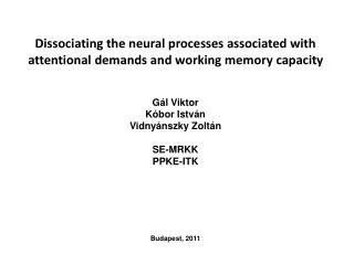
Dissociating the neural processes associated with attentional demands and working memory capacity. Gál Viktor Kóbor István Vidnyánszky Zolt á n SE-MRKK PPKE-ITK Budapest, 2011. Measuring the neural effect of working memory load using fMRI . .

E N D
Dissociating the neural processes associated with attentional demands and working memory capacity Gál Viktor Kóbor István VidnyánszkyZoltánSE-MRKK PPKE-ITK Budapest, 2011
Measuring the neural effect of working memory load using fMRI. • Working memory is affected by several CNS disorders • WM tasks (e.g. delayed match to sample) are important part of neuropsychatric tests • The goal is to show that fMRI can be used to measure: • -Subtle changes in the neural responses in specific brain regions associated with memory-load dependent modulation of the performance in a working memory tasks. • -Neural correlates of the individual differences in WM capacity.
VSTM experiment • 13 subjects, 3 excluded(poor behavioral performance and scanning artifacts) • Plan: to have at least 20-25 subjects in the analysis • Main experiment + independent ROI localizer scan • Standard GLM analysis protocols
fMRI measurements, analysis • Scanning parameters: • Philips Achieva 3T, 36 functional axial slices, TR: 2 s, resolution: 3 x 3 x 3.5mm. • Preprocessing, analysis: • Functional data was preprocessed using standard procedures in SPM8. • Data was analyzed by standard GLM procedures via customized SPM8 scripts. • Statistical parametric maps were transformed to the MNI space using thetransformation parameters of the normalized high resolution T1W anatomical scans.
Behavioural results where K is the memory capacity, S is the size of the array, H is the observed hit rate and F is the false alarm ra
Localization of VSTM related BOLD activity Random effect group results of the memory load 5 > baselinecontrast map group-wise analysis at a threshold of FDR=0.05.
Localization of VSTM related BOLD modulation Random effect group results of the memory load 5 > memory load 1 contrast map group-wise analysis at a threshold of p<0.001.
Correlation of bold responses and behavioral performance • Correlation between the memory load induced modulation of bold responses and behavioral performance R=0.64, p=0.048
Conclusion • It was found that fMRI responses: • in the visual cortex showed early saturation: increasing the number of objects tobe remembered above two resulted in a very small modulation of the BOLD responses in these regions • in the bilateral supplemental motor area extending into middle cingulatedcortex and the right anterior insula gradually increased with increasing the WMload, independently of the subjects’ WM capacity. • in the bilateral intraparietal sulcus and in the left anterior insula closely associatedwith the subjects’ WM capacity and not with the overall attentional demands. • To conclude the results provide evidence that different neural networks areinvolved in the processes associated with WM capacity and changes in overall attentional demands
Functional connectivity magnetic resonance imaging (fcMRI) RestingstatefcMRI (rs-fcMRI) studies measure the correlations in spontaneous activity between brain regions . These rs-fcMRI measurements are reliable across scans and institutions and are thought to have been shaped by the cumulative effect of experiences across one’s lifespan. Important group-level rs-fcMRI studies have already shown differences in spontaneous activity in disorders such as autism, schizophrenia, depression, and attention-deficit hyperactivity disorder.
fcMRI parameters, preprocessing • 11 subjects, 15min • Scanning parameters: • Philips Achieva 3T, 28 functional axial slices, TR: 1.5 s, resolution: 3 x 3 x 4mm and 36 functional axial slices, TR: 2 s, resolution: 3 x 3 x 3.5mm • Preprocessing, analysis: • Functional data was preprocessed using standard procedures in SPM8. • Noise (artificial correlation) was attenuated by: • Regressing out • Motion correction parameters • Phys. monitoring data (respiration, pulsoxy) • White matter signal • Global gray matter signal • Bandpass filter: [0.009Hz 0.08Hz]
fcMRI parameters, preprocessing • Seed ROI: left and right insula anterior based on main memory experiment group results • backprojected from MNI to single subject space • Correlation between seed voxels and all other gray matter voxels were calculated and entered in second level analysis • Group level statistical parametric maps werecreated after random effects analysis of the individual SPMs


![Attention, Working Memory, and Executive Function [Processes Under Construction]](https://cdn2.slideserve.com/4587086/slide1-dt.jpg)