Download
1 / 64
640 likes | 806 Views
Introduction and Tissues. Human Anatomy BIOL 1010 Liston Campus. What is Anatomy?. Anatomy (= morphology): study of body’s structure Physiology: study of body’s function Structure reflects Function!!! Branches of Anatomy Gross: Large structures Surface: Landmarks

E N D
Introduction and Tissues Human AnatomyBIOL 1010 Liston Campus
What is Anatomy? • Anatomy (= morphology): study of body’s structure • Physiology: study of body’s function • Structure reflects Function!!! • Branches of Anatomy • Gross: Large structures • Surface: Landmarks • Histology: Cells and Tissues • Developmental: Structures change through life • Embryology: Structures form and develop before birth
Hierarchy of Structural Organization Each of these build upon one another to make up the next level: • Chemical level • Cellular • Tissue • Organ • Organ system • Organism
Hierarchy of Structural Organization • Chemical level • Atoms combine to make molecules • 4 macromolecules in the body • Carbohydrates • Lipids • Proteins • Nucleic acids
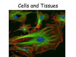
Hierarchy of Structural Organization • Cellular • Made up of cells and cellular organelles (molecules) • Cells can be eukaryotic or prokaryotic • Organelles are structures within cells that perform dedicated functions (“small organs”) http://cmweb.pvschools.net/~bbecke/newell/Cells.html
Hierarchy of Structural Organization • Tissue • Collection of cells that work together to perform a specialized function • 4 basic types of tissue in the human body: • Epithelium • Connective tissue • Muscle tissue • Nervous tissue www.emc.maricopa.edu
Hierarchy of Structural Organization • Organ • Made up of tissue • Heart • Brain • Liver • Pancreas, etc…… Pg 181
Hierarchy of Structural Organization • Organ system (11) • Made up of a group of related organs that work together • Integumentary • Skeletal • Muscular • Nervous • Endocrine • Cardiovascular • Lymphatic • Respiratory • Digestive • Urinary • Reproductive Circulatory Pg 341 Urinary System
Hierarchy of Structural Organization • Organism • An individual human, animal, plant, etc…… • Made up all of the organ systems • Work together to sustain life
Anatomical Directions • Anatomical position • Regions • Axial vs. Appendicular • Anatomical Directions-It’s all Relative! • Anterior (ventral) vs. Posterior (dorsal) • Medial vs. Lateral • Superior (cranial) vs. Inferior (caudal) • Superficial vs. Deep • Proximal vs. Distal • Anatomical Planes • Frontal = Coronal • Transverse = Horizontal = Cross Section • Sagittal Pg 5
4 Types of Tissue • Epithelium • Connective • Muscle • Nervous
Tissues: groups of cells closely associated that have a similar structure and perform a related function • Four types of tissue • Epithelial = covering/lining • Connective = support • Muscle = movement • Nervous = control • Most organs contain all 4 types • Tissue has non-living extracellular material between its cells
EPITHELIAL TISSUE: sheets of cells cover a surface or line a cavity • Functions • Protection • Secretion • Absorption • Ion Transport
Characteristics of Epithelium • Cellularity • Composed of cells • Specialized contacts • Joined by cell junctions • Polarity • Apical vs. Basal surfaces differ • Supported by connective tissue • Avascular • Innervated • Highly regenerative
Classification of Epithelium-based on number of layers and cell shape • Layers • Simple • Stratified • Stratified layers characterized by shape of apical layer • Psuedostratified • Shapes • Squamous • Cuboidal • Columnar • Transitional
Simple squamous (1 layer) Lungs, blood vessels, ventral body cavity Simple cuboidal Kidney tubules, glands Simple columnar Stomach, intestines Pseudostratified columnar Respiratory passages (ciliated version) Stratified squamous (>1 layer) Epidermis, mouth, esophagus, vagina Named so according to apical cell shape Regenerate from below Deep layers cuboidal and columnar Transitional (not shown) Thins when stretches Hollow urinary organs Types of Epithelium All histology pictures property of BIOL 1010 Lab
Endothelium Simple squamous epithelium that lines vessels e.g. lymphatic & blood vessel Mesothelium Simple squamous epithelium that forms the lining of body cavities e.g. pleura, pericardium, peritoneum Special Epithelium
Features of Apical Surface of Epithelium • Microvilli:(ex) in small intestine • Finger-like extensions of the plasma membrane of apical epithelial cell • Increase surface area for absorption • Cilia: (ex) respiratory tubes • Whip-like, motile extension of plasma membrane • Moves mucus, etc. over epithelial surface 1-way
Features of Lateral Surface of Epithelium • Cells are connected to neighboring cells via: • Contour of cells-wavy contour fits together • Cell Junctions (3 common) • Desmosomes • Proteins hold cells together to maintain integrity of tissue • Tight Junctions • Plasma membrane of adjacent cells fuse, nothing passes • Gap junction • Proteins allow small molecules to pass through
Features of the Basal Surface of Epithelium • Basement membrane • Sheet between the epithelial and connective tissue layers • Attaches epithelium to connective tissue below • Made up of: • Basal lamina: thin, non-cellular, supportive sheet made of proteins • Superficial layer • Acts as a selective filter • Assists epithelial cell regeneration by moving new cells • Reticular fiber layer • Deeper layer • Support
Glands • Epithelial cells that make and secrete a product • Products are water-based and usually contain proteins • Classified as: • Unicellular vs. multicellular • Exocrine vs. Endocrine Page 138
Glands: epithelial cells that make and secrete a water-based substance w/proteins • Exocrine Glands • Secrete substance onto body surface or into body cavity • Activity is local • Have ducts • Unicellular or Multicellular • (ex) goblet cells, salivary, mammary, pancreas, liver
Glands: epithelial cells that make and secrete a water-based substance w/proteins • Endocrine Glands • Secrete product into blood stream • Either stored in secretory cells or in follicle surrounded by secretory cells • Hormones travel to target organ to increase response (excitatory) • No ducts • (ex) pancreas, adrenal, pituitary, thyroid
4 Types of Tissue • Epithelium • Connective • Muscle • Nervous
4 Types of Connective Tissue Connective Tissue Proper Cartilage Bone Tissue Blood
Connective Tissue (CT): most abundant and diverse tissue • Four Classes • Functions include connecting, storing & carrying nutrients, protection, fight infection • CT contains large amounts of non-living extracellular matrix • Contains a variety of cells and fibers • Some types vascularized • All CT originates from mesenchyme • Embryonic connective tissue
Fibers in Connective Tissue • Fibers For Support • Reticular: • form networks for structure & support • (ex) cover capillaries • Collagen: • strongest, most numerous, provide tensile strength • (ex) dominant fiber in ligaments • Elastic: • long + thin, stretch and retain shape • (ex) dominant fiber in elastic cartilage
Components of Connective Tissue • Fibroblasts: • cells that produce all fibers in CT • produce + secrete protein subunits to make them • produce ground matrix • Interstitial (Tissue) Fluid • derived from blood in CT proper • medium for nutrients, waste + oxygen to travel to cells • found in ground matrix • Ground Matrix (substance): • part of extra-cellular material that holds and absorbs interstitial fluid • Made and secreted by fibroblasts • jelly-like with sugar & protein molecules
1) Connective Tissue Proper • Two kinds: Loose CT & Dense CT • Functions • Support and bind to other tissue • Hold body fluids • Defends against infection • Stores nutrients as fat • Each function performed by different kind of fibers and cells in specific tissue
Defense from Infection • Areolar tissue below epithelium is body’s first defense • Cells travel to CT in blood • Macrophages-eat foreign particles • Plasma cells-secrete antibodies, mark molecules for destruction • Mast cells-contain chemical mediators for inflammation response • White Blood Cells = neutrophils, lymphocytes, eosinophils-fight infection • Ground substance + cell fibers-slow invading microorganisms
Areolar CT All types of fibers present All typical cell types present Surrounds blood vessels and nerves Loose CT Proper
Adipose tissue Loaded with adipocytes, highly vascularized, high metabolic activity Insulates, produces energy, supports Found in hypodermis under skin Reticular CT Contains only reticular fibers Forms caverns to hold free cells, forms internal “skeleton” of some organs Found in bone marrow, holds blood cells, lymph nodes, spleen Specialized Loose CT Proper
Dense/Fibrous Connective Tissue • Contains more collagen • Can resist extremely strong pulling forces • Regular vs. Irregular • Regular-fibers run same direction, parallel to pull • (eg) fascia, tendons, ligaments • Irregular-fibers thicker, run in different directions • (eg) dermis, fibrous capsules at ends of bones Denseregular Denseirregular
2) Cartilage • Chondroblastsproduce cartilage • Chondrocytes mature cartilage cells • Reside in lacunae • More abundant in embryo than adult • Firm, Flexible • Resists compression • (eg) trachea, meniscus • Avascular (chondrocytes can function w/ low oxygen) • NOT Innervated • Perichondrium • dense, irregular connective tissue around cartilage • growth/repair of cartilage • resists expansion during compression of cartilage
Cartilage in the Body • Three types: • Hyaline • most abundant • fibers in matrix • support via flexibility/resilience • (eg) at limb joints, ribs, nose • Elastic • many elastic fibers in matrix too • great flexibility • (eg) external ear, epiglottis • Fibrocartilage • resists both compression and tension • (eg) meniscus, annulus fibrosus
3) Bone Tissue: (a bone is an organ) • Well-vascularized • Function: • support (eg) pelvic bowl, legs • protect (eg) skull, vertebrae • mineral storage (eg) calcium, phosphate (inorganic component) • movement (eg) walk, grasp objects • blood-cell formation (eg) red bone marrow
Bone Tissue • Osteoblasts • Secrete organic part of bone matrix • Osteocytes • Mature bone cells • Sit in lacunae • Maintain bone matrix • Osteoclasts • Degrade and reabsorb bone • Periosteum • External layer of CT that surrounds bone • Outer: Dense irregular CT • Inner: Osteoblasts, osteoclasts • Endosteum • Internal layer of CT that lines cavities and covers trabeculae • Contains osteoblasts and osteoclasts academic.kellogg.cc.mi.us/.../skeletal.htm
External layer Osteon (Haversian system) Parallel to the long axis of the bone Groups of concentric tubules (lamella) Lamella = layer of bone matrix where all fibers run in the same direction Adjacent lamella fibers run in opposite directions Haversian Canal runs through center of osteon Contains blood vessels and nerves Connected to each other by perforating (Volkman) canals Interstitial lamellae fills spaces and forms periphery Compact Bone www.mc.vanderbilt.edu/.../CartilageandBone03.htm
Bone Anatomy: Spongy bone • Spongy bone (cancellous bone): internal layer • Trabeculae: small, needle-like pieces of bone form honeycomb • each made of several layers of lamellae + osteocytes • no canal for vessels • space filled with bone marrow • not as dense, no direct stress at bone’s center
Shapes of Bones • Flat = skull, sternum, clavicle • Irregular = pelvis, vertebrae • Short = carpals, patella • Long = femur, phalanges, metacarpals, humerus
Anatomy of a Long Bone • Diaphysis • Medullary Cavity • Nutrient Artery & Vein • 2 Epiphyses • Epiphyseal Plates • Epiphyseal Artery & Vein • Periosteum • Does not cover epiphyses • Endosteum • Covers trabeculae of spongy bone • Lines medullary cavity of long bones training.seer.cancer.gov/.../illu_long_bone.jpg
2 Types of Bone Formation • Intramembranous Ossification • Membrane bones: most skull bones and clavicle • Osteoblasts in membrane secrete osteoid that mineralizes • Endochondral Ossification: All other bones • Begins with a cartilaginous model • Cartilage calcifies • Medullary cavity is formed by action of osteoclasts • Epiphyses grow and eventually calcify • Epiphyseal plates remain cartilage for up to 20 years
Bone Growth & Remodeling • GROWTH • Appositional Growth = widening of bone • Bone tissue added on surface by osteoblasts of periosteum • Medullary cavity maintained by osteoclasts • Lengthening of Bone • Epiphyseal plates enlarge by chondroblasts • Matrix calcifies (chondrocytes die and disintegrate) • Bone tissue replaces cartilage on diaphysis side • REMODELING • Due to mechanical stresses on bones, their tissue needs to be replaced • Osteoclasts-take up bone ( = breakdown) release Ca2++ , PO4 to body fluids from bone • Osteoblasts-form new bone by secreting osteoid • Ideally osteoclasts& osteoblastswork at the same rate!
4) Blood: Atypical Connective Tissue • Function: • Transports waste, gases, nutrients, hormones through cardiovascular system • Helps regulate body temperature • Protects body by fighting infection • Derived from mesenchyme • Hematopoiesis: production of blood cells • Occurs in red bone marrow • In adults, axial skeleton, girdles, proximal epiphyses of humerus and femur
Blood Cells • Erythrocytes: (RBC) small, oxygen-transporting • most abundant in blood • no organelles, filled w/hemoglobin • pick up O2 at lungs, transport to rest of body • Leukocytes: (WBC) complete cells , 5 types • fight against infectious microorganisms • stored in bone marrow for emergencies • *Platelets = Thrombocytes: • fragments of cytoplasm • plug small tears in vessel walls, initiates clotting