Chapter 28
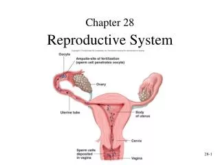
Chapter 28. Reproductive System. Sperm Cell Development.

Chapter 28
E N D
Presentation Transcript
Chapter 28 Reproductive System
Sperm Cell Development • Spermatozoa produced in seminiferous tubules. At puberty, GnRH (Gonadotropin-releasing hormone) secretion increases leading to increased LH and FSH release from anterior pituitary. (Note: GnRH is synthesized and released from neurons within the hypothalamus) FSH levels promote sperm formation, FSH levels promote interstitial cells to produce large amounts of testosterone. • Spermatogonia (germ cell) divide (mitosis) to form primary spermatocytes and daughter spermatagonia • Primary spermatocytes divide (first division of meiosis) to form secondary spermatocytes • Secondary spermatocytes divide (second division of meiosis) to form spermatids
Sperm Cell Development • Spermatids develop an acrosome (helps sperm penetrate ovum) and flagellum • Sustentacular (Sertoli, nurse) cells nourish sperm cells and form a blood-testis barrier(isolates sperm from immune system) and produce hormones (androgens, estrogens, & inhibins) • Interstitial cells produce testosterone. Sustentacular cells convert it to dihydrotestosterone (DHT) and estrogen. These are active hormones that promote sperm formation
Puberty • Before birth, placenta secretes human chorionic gonadotropin hormone which stimulates secretion of testosterone by fetal testes (testosterone causes the enlargement & differentiation of male genitals & is required for descent of testes near end of fetal development) • From birth to puberty, no stimulation of secretion of testosterone • Puberty: age at which individuals become capable of sexual reproduction • Previous to puberty: small amounts of testosterone from adrenal gland inhibits GnRH • At puberty, pituitary becomes less sensitive to testosterone inhibition. Amount of GnRH increases, amount of LH and FSH increases • Elevated FSH causes sperm cell formation • Elevated LH causes interstitial cells to secrete larger amounts of testosterone
Maturation and Fertilization of Oocyte • Oogenesis is the production of a secondary oocyte in ovaries • Oogonia(stem cell) are cells from which oocytes develop. The oogonia divide by mitosis to produce other oogonia and primary oocytes. • Five million oocytes produced by the 4th month of prenatal life. At birth, about 2 million begin first meiotic division but stop at prophase (at this stage called primary oocyte). All remain at this state until puberty. • Note: oogonia can form after birth from stem cells but unclear. • Primary oocytes are surrounded by granulosa cells and called a primordial follicle • Primordial follicle becomes a primary follicle when oocyte and granular cells enlarge (usually occurs at puberty) • Primary follicle becomes secondary follicle (which is released during ovulation) and enlarges to form mature or Graafian follicle • Usually only one is ovulated, others degenerate
Ovulation, Fertilization, Follicle Fate • Ovulation: release of a secondary oocyte from an ovary. Unlike spermatogenesis, division of cytoplasm during meiosis is uneven and polar bodies are very small, oocyte very large • Graafian follicle become corpus luteum (endocrine body that secretes progesterone & estrogen) • Fertilization: begins when a sperm cell binds the plasma membrane of secondary oocytes and penetrates into cytoplasm. • Secondary oocyte completes meiosis II forming one polar body. Fertilized egg now a zygote • Fate of corpus luteum • If fertilization occurs, corpus luteum persists • If no fertilization, becomes corpus albicans (atrophied corpus luteum)
Menstrual Cycle • Puberty • Begins with menarche (first episode of menstrual bleeding) • Begins when GnRH levels increase • Menstrual Cycle • About 28 days long • Phases • Menses • Proliferative or follicular phase (functional layer proliferates; follicle matures) (time between the ending of menses & ovulation) • Secretory or luteal phase (maturation and secretion of uterine glands; presence of corpus luteum) (period after ovulation & before next menses) • Menses • Amenorrhea: absence of a menstrual cycle • Menopause: cessation of menstrual cycles
Female fertility • Sperm ejaculated into vagina during copulation and transported through cervix and uterine tubes to ampulla • Sperm cells undergo capacitation, enabling them to release acrosomal enzymes to digest away follicular cells • Pregnancy • Oocyte can be fertilized up to 24 hours after ovulation • Sperm cells can be viable for up to 6 days in female tract • Ectopic pregnancy: Implantation occurs anywhere other than uterine cavity • Fertilization. Occurs in uterine tube. Multiple mitoses occur after union of oocyte and sperm nuclei, (pronuclei) forming an embryo. Outer layer of embryonic mass is the trophoblast. Implantation. Trophoblast also secretes HCG (keeps corpus luteum from degenerating; blood levels of estrogen & progesterone do not decrease & menses does not occur).
Blastocyst Formation • Zygote divides to form 2 cells about 18-39 hours after fertilization • 2 cells divide to form 4, 8, and so on • Pluripotent: Ability to develop into wide range of tissues • Morula: solid ball of 12 or more cells • Blastocyst or hollow sphere of cells • Implantation: burrowing into uterine wall • Placenta develops from trophoblast cells
Formation of Placenta • Frontal section of the uterus and uterine tube showing development 7 days after fertilization • Implantation of the blastocyst with syncytiotrophoblast beginning to invade the uterine wall at 8-12 days
Placenta Formation • Intermediate stage of placental formation at about 14-20 days. As maternal blood vessels are encountered by the syncytiotrophoblast, lacunae are formed and filled with maternal blood • Cytotrophoblast cords surround the syncytiotrophoblast and lacunae, and embryonicblood vessels enter the cord at about 1 month
Mature Placenta and Fetus ( fetal blood vessels & maternal blood vessels are in close contact, & nutrients are exchanged between fetal & maternal blood, but fetal & maternal blood do not mix)
Formation of the Germ Layers • Amniotic cavity: forms inside inner cell mass and surrounded by layer of cells called the amnion or amniotic sac. Forms after implantation • Embryonic disk: composed of ectoderm and endoderm and is part of the inner cell mass • Amniotic cavity eventually surrounds the developing embryo providing a protective fluid bag • Yolk sac: forms inside blastocele from endoderm & provides nutrition to the developing embryo.