Download
1 / 24
270 likes | 646 Views
Neuroimaging of Toxic Encephalopathy. Ecstasy. Sandy CY Chen, MD Tri-Service General Hospital, Taiwan. SNR 2010 Bolongna. Toxic Encephalopathy. Etiology Pathomechanisms Imaging approach By etiologies: drugs By history Substance abuse: imaging patterns

E N D
Neuroimaging of Toxic Encephalopathy Ecstasy Sandy CY Chen, MD Tri-Service General Hospital, Taiwan SNR 2010 Bolongna
Toxic Encephalopathy • Etiology • Pathomechanisms • Imaging approach By etiologies: drugs By history • Substance abuse: imaging patterns • Occupational & environmental • Suicidal attempts
Toxic Encephalopathy -Pathomechanisms • Leukoencephalopathy: demyelination, ischemia and astrogliosis • Excitotoxicity • Vasculopathy • Cellular energy failure
Glutamate Excitotoxicity Cellular edema Apoptosis or necrotic cell death Moritani et al. AJNR 2005
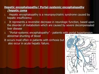
Hyperammonemia induce low grade brain edema by impairing glutamate-nitric oxide-cGMP pathway
Hyperammonemia In Hepatic encephalopathy • Impair glutamate-nitric oxide-cGMP pathway • Disrupt osmolar balance: glutamine mI & Choline NAA Cr GLX mI Cho Chronic relapsing hepatic encephalopathy
Excitotoxicity in Wernicke’s encephalopathy Acetyl CoA Krebs cycle Glutamate α-Ketoglutarate α-KGDH Succinyl CoA Succinate
NMDA –mediated excitotoxicity In Wernicke Encephalopathy Thiamine-dependent enzyme α-ketoglutarate dehydrogenase
Serotonergic pathway Serotonergic : Memory, mood, cognitive and motor NEJM 2005
Ecstasy encephalopathy High density of 5HT Receptors in globus pallidus Oxidative stress, excitotoxicity, and mitochondrial dysfunction
+ + () () - - - - - ( ) ( ) ( ) ( ) ( ) Basal ganglia-thalamocortical circuit ← MDMA effect Disinhibition
To investigate the effects of MDMA abuse on the alterations of brain metabolites and anatomical connectivity related to the function of the basal ganglia-thalamocortical circuit using 1H-MRS and DTI Study purpose
Materials and Methods 31 MDMA (24 male, 7 female, aged 18 - 34) abusers with positive hair and urine tests were enrolled as MDMA group, and 33 normal healthy non-abusers (17 male, 16 female, aged 21 - 32) were also included as controls. MR images were acquired at a 3.0 T MR system (Philips, Achieva, Best, Netherlands).
LCModel analysis for MRS Single voxel proton MRS 3T scanner Double spin-echo, point resolved spectroscopy (PRESS) TR/TE 2000/40 ms, NEX=128
DTI analysis Diffusion tensor imaging • Diffusion tensor imaging were acquired with 6 non-collinear directions with b =1000s/mm2. -- TR/TE=5890/57ms, matrix size=128x128, slice thickness=3 mmwithout gap, NEX=6, number of slice=40. • The FA difference was statistically compared on a voxel-by-voxel basis between normal and MDMA groups using Student’s t-test.
Whole brain FA images: normalized to anICBM-MNI-FA template using affine registration followed by diffeomorphic demons registration DTI ( MDMA group ) ( control group ) #1 #9 #10 #28 #10 #1 #9 #28 … … … … Average brain Average brain
Results MDMA > control MDMA < control voxel-wise P < 0.05 cluster > 50 voxels MDMA < control MDMA > control
Discussion • The increased mI in the basal ganglia raise the possibility that MDMA-induced deficits on the basal ganglia would reduce inhibitory effects on the thalamus, and therefore increase the thalamocortical activities
Discussion • The observed ADC reduction in the corticospinal tracts and thalamus in the present study would likely reflect the increased neuronal activity in the thalamorcortical fibers
Discussion • Study limitations 1) many MDMA users have also exposed to other recreational drugs. 2) the causative relationships between MDMA use and the alternations in the basal-thalamocortical circuit can be challenged because we did not conduct standard motor function assessments in MDMA subjects
+ + () () - - - - - ( ) ( ) ( ) ( ) ( ) Conclusion mI ← MDMA effect Disinhibition Basal ganglia-thalamocortical circuit
Contributions: Professor Hsiao-Wen Chung Ming-Chung Chou Ph.D. Nei-Yu Chuo Hua-Shan Liu Chao-In Wang M.D. Hueng-Wen Gao Chun-Jung Juan Chun-Jen Hsueh *National Taiwan University *National Defense Medical Center *Tri-Service General Hospital 0