Download
1 / 1
10 likes | 146 Views
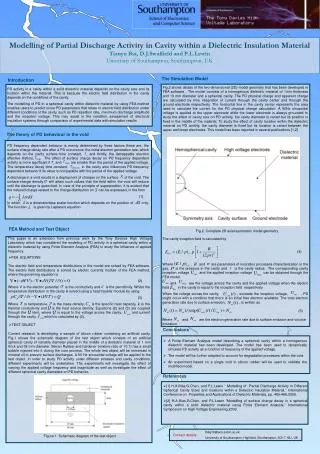
Proposed method. Introduction. In the last decade, technological improvements of radiotherapy (RT) hardware and software have been significant and consequently the use and importance of RT in cancer treatment have increased greatly.

E N D
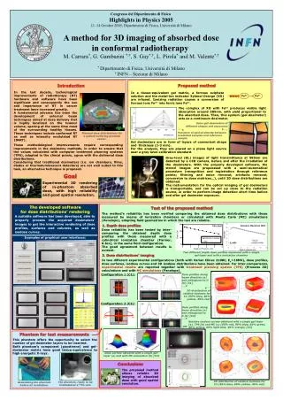
Proposed method Introduction In the last decade, technological improvements of radiotherapy (RT) hardware and software have been significant and consequently the use and importance of RT in cancer treatment have increased greatly. A fundamental advance has been the development of external beam techniques aimed at dose delivery that is highly localized on the tumour volume, sparing at the same time most of the surrounding healthy tissues. These techniques include conformal RT as well as intensity modulated RT (IMRT). In a tissue-equivalent gel matrix, a ferrous sulphate solution and the metal-ion indicator Xylenol Orange (XO) are infused. Ionizing radiation causes a conversion of ferrous ions Fe2+ into ferric ions Fe3+. The complex of XO with Fe3+ produces visible light absorption around 585nm, with yield proportional to the absorbed dose. Then, this system (gel-dosimeter) acts as a continuum dosimeter. Fe2+ Fe3+ RADIATION Some gel-dosimeters of different shapes and exposures Variation of optical density between irradiated samples and reference sample Planned dose distributions for a patient suffering prostate cancer. Gel dosimeters are in form of layers of convenient shape and thickness (1-3 mm). For the analysis, they are placed on a plane light source near a grey-level calibration standard. These methodological improvements require corresponding improvements in the dosimetry methods, in order to ensure that the values calculated with computer treatment planning systems (TPS), adopted in the clincal praxis, agree with the delivered dose distributions. Considering that traditional dosimeters (i.e. ion chambers, films, diodes or thermoluminescent detectors) are not well suited to this task, an alternative technique is proposed. Grey-level (GL) images of light transmittance at 585nm are detected by a CCD camera, before and after the irradiation of the dosimeters. With the properly developed software, the stored images are propcessed with a semi-automatical procedure (recognition and registration through reference points; filtering and noise removal; artefacts removal; conversion to dose matrices…), until 3D dose distributions are obtained. The instrumentation for the optical imaging of gel dosimeters is transportable, and can be set up close to the radiation source, in order to perform image detection short time before and after gel dosimeter exposure. Goal Experimental 3D rendering of in-phantom absorbed dose, with high reliability and good spatial resolution. CCD Controller The developed software for dose distributions’ rendering Test of the proposed method CCD Filter 585nm The method’s reliability has been verified comparing the obtained dose distributions with those measured by means of ionization chambers or calculated with Monte Carlo (MC) simulations (Penelope), adopting field geometries in which the last are reliable. A suitable software has been developed, able to properly process the acquired dosimeters’ images to get the interactive rendering of dose profiles, surfaces and volumes, as well as isodose curves. gel 1. Depth dose profiles Illuminator Siemens Mevatron MX2 60Co Unit Dose reliability has been tested by inter-comparing the obtained depth dose profiles with those measured with a cylindrical ionization chamber (Farmer, 0.6cc), in the same field configuration. The good agreement between results is evident. Examples of graphical user interfaces: Eγ=6MV Two different depth dose profiles measured with a single gel-layer and with a ionization chamber 2. Dose distributions’ imaging In two different experimental configurations (both with Varian Clinac 2100C, Eγ=18MV), dose profiles, dose surfaces, isodose curves and 3D isodose distributions have been obtained. For inter-comparisons, experimental results are reported together with treatment planning system (TPS) (Prowess 3D) calculations and with MC simulations (Penelope). Configuration 1 (C1): Dose profiles along beam direction (a) and orthogonal to it (b) [C1] gel layers gel layers (b) 1 1 2 2 3 3 4 4 5 5 6 6 90° 3x2 field 3x2 field (a) phantom phantom 3D distribution of relative isodoses for C1 (95% blue, 80% yellow, 40% red) Configuration 2 (C2): (b) (a) Dose profiles along beam direction (a) and orthogonal to it (b) [C2] 270° Relative isodose curves obtained with a single gel-layer (a), TPS (b) and MC (c) (95% red, 90% blue, 85% green, 80% yellow, 60% light blue, 40% orange) [C2] Phantom for test measurements This phantom offers the opportunity to select the number of gel dosimeter layers to be inserted. Both phantom’s component (poystirene) and gel-dosimeter matrix have good tissue-equivalence to high energetic X-rays. (a) (a) (b) Dose surface obtained with a single gel-layer (a) and with MC simulation (b) [C2] Conclusions (b) The proposed method allows reliable 3D imaging of absorbed dose with good spatial resolution. 3D distribution of relative isodoses for C2 (95% blue, 80% yellow, 40% red) The phantom, ready to be irradiated at a 60Co unit. Assembling the phantom before its irradiation. (c) Congresso del Dipartimento di Fisica Highlights in Physics 2005 11–14 October 2005, Dipartimento di Fisica, Università di Milano A method for 3D imaging of absorbed dose in conformal radiotherapy M. Carrara*, G. Gambarini *,†, S. Gay*,†, L. Pirola* and M. Valente*,† * Dipartimento di Fisica, Università di Milano † INFN – Sezione di Milano