1 / 25
0 likes | 126 Views
Disseminated intravascular coagulation (DIC) is an acquired syndrome characterized by excessive systemic activation of coagulation, resulting in both hemorrhage and thrombosis. <br>OR<br>Is a condition in which blood clots form throughout the body, blocking small blood vessels<br><br>DIC can progress rapidly into life-threatening multiorgan failure; thus, identifying the underlying etiology is paramount to management

E N D
DISSEMINATED INTRAVASCULAR COAGULATION (DIC) Dr Beatrice kyomugisa Paediatrician Fort portal RRH
Introduction/Definition • Disseminated intravascular coagulation (DIC) is an acquired syndrome characterized by excessive systemic activation of coagulation, resulting in both hemorrhage and thrombosis. OR • Is a condition in which blood clots form throughout the body, blocking small blood vessels • DIC can progress rapidly into life-threatening multiorgan failure; thus, identifying the underlying etiology is paramount to management
Epidemiology • DIC may occur in 30-50% of patients with sepsis • It develops in an estimated 1% of all hospitalized patients • DIC occurs at all ages and in all races, and no particular sex predisposition has been noted.
Causes of disseminated intravascular coagulation in children 1. Infection • Bacteria: Meningococcus, gram-positive and -negative bacterial sepsis • Virus: HIV, varicella-zoster, cytomegalovirus, Dengue fever, Ebola virus • Fungal: Candida, Aspergillus • Rickettsia : Rocky Mountain Spotted fever • Parasites: Malaria 2. Injury • Brain injury • Crush injury • Massive burns • Extensive surgery 3. Malignancy • Acute promyelocytic leukemia • Acute lymphoblastic leukemia 4. Microangiopathic disorders • Giant hemangioma – Kasabach-Merritt syndrome
Cont….. 5. Neonatal causes • Birth asphyxia • Respiratory distress syndrome • Meconium aspiration • Amniotic fluid aspiration • Necrotizing enterocolitis • Congenital infections: Neonatal cytomegalovirus, herpes simplex virus, bacterial or fungal infections 6. Gastrointestinal disease • Acute and chronic liver disease • Reye syndrome 7. Congenital thrombotic disorders • Homozygous deficiencies of proteins C and S • Antithrombin III deficiency
Risk factors of DIC in neonates • Extreme prematurity • Extremely low birth weight • Low APGAR scores
Why is DIC common in neonates • Platelets and coagulation factors are first detectable at 5 and 10 weeks of gestation, respectively. • Platelets reach normal range of 150–450×109 /L at 22 weeks of gestation and most coagulation factors achieve adult values by 6 months of postnatal age, with a few notable exceptions. • Platelets are found to be hyporeactive and aggregation is impaired due to deficiency of α-adrenergic receptors on platelet membrane, especially in preterm infants
Stages of DIC Stage 1: Overactive clotting leads to formation of blood clots throughout the blood vessels. The clots can reduce or block blood flow, which can damage vital organs. Stage 2: As DIC progresses, the overactive clotting uses up platelets and clotting factors that help the blood to clot. Without these platelets and clotting factors, DIC leads to bleeding just beneath the skin, in the nose or mouth, or deeper tissues.
Pathogenesis of DIC Intravascular activation of coagulation: • Tissue damage from the initiating underlying disease releases procoagulants into the bloodstream, resulting in intravascular activation of coagulation. Examples of procoagulants include lipopolysaccharides from bacteria, phospholipids from damaged vascular endothelium, or the formation of neutrophil extracellular traps. Formation of fibrin in the circulation: • Tissue procoagulants activate hemostasis primarily through the interaction of tissue factor and factor VII, thereby promoting fibrin formation and deposition within the microcirculation.
Cont…. Fibrinolysis • Fibrin formation activates the fibrinolytic pathway, which produces plasmin that cleaves fibrinogen and fibrin, thereby generating fibrin degradation products (FDPs). FDPs interfere with fibrin polymerization and impair platelet aggregation. Consumption of clotting factors and platelets: • Ongoing activation of the coagulation system and fibrin deposition consume clotting factors and platelets.
Cont…. Hemolysis: • Intravascular fibrin strands cause mechanical shearing of red blood cells, resulting in microangiopathic hemolytic anemia. End-organ damage: • Deposition of fibrin into the microcirculation of organs results in tissue ischemia and damage.
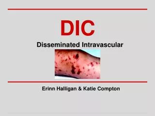
Clinical features of DIC • Bleeding venipuncture sites • Oozing of blood from indwelling catheters • Spontaneous or minimal trauma-related generalized ecchymoses • Development of large, bullous hemorrhagic skin lesions on previous viral exanthematous sites • Mucosal bleeding from gingiva, gastrointestinal or renal tracts • Unexpected and major bleeding around drain sites or surgical wounds post surgery in postoperative states • Thrombophlebitis at unusual sites • Renal impairment in the absence of other explanations • Fluctuating central nervous system disturbances like confusion, and seizures—consistent with the microcirculatory ischemia • Respiratory distress syndrome with no obvious explanation • Dermal infarcts and skin necrosis • Greyish discoloration of finger tips, toes or ear lobes, which has been termed ‘acral cyanosis’; usually seen in extreme cases
Complications of DIC • Multi organ failure e.g acute renal failure • Life threatening hemorrhage e.g. Pulmonary embolism and hemorrhage • Gangrene • Venous thromboembolism • Shock • Stroke • Acute respiratory distress syndrome
Differential diagnosis • Dysfibrinogenemia • Hemolytic uremic syndrome • Heparin-induced thrombocytopenia • Immune thrombocytopenia (ITP) • Thrombotic thrombocytopenic purpura (TTP) • Hepatic failure • Vit K deficiency
Peripheral smear in microangiopathic hemolytic anemia showing presence of schistocytes • Multiple helmet cells (arrows) • Fragmented red cells (small arrowhead) • Microspherocytes are also seen (large arrowheads). • The platelet number is reduced; the large platelet in the center (dashed arrow) suggests that the thrombocytopenia is due to enhanced destruction.
Diagnosis Consider the diagnosis of DIC if all the following criteria are met: • The patient has an underlying condition that predisposes them to DIC (eg, infection, trauma, malignancy) • Laboratory testing is consistent with DIC (ie, thrombocytopenia plus coagulation factor consumption [prolonged PT and aPTT, low fibrinogen] and fibrinolysis [elevated D-dimer]) • No other etiology for these findings has been identified • Clinical evidence of bleeding or thrombosis are supportive of the diagnosis but are not required for diagnosis.
Management • Treat an underlying condition that predisposes them to DIC (eg, infection, trauma, malignancy). Antibiotics for severe sepsis • Transfusion of platelets (10 to 15mls/kg) • Replacement of clotting factors: Clotting factors can be replaced by either FFP or cryoprecipitate. Repeat transfusions may be necessary. FFP dose of 10 to 15 mL/kg per infusion. Cryoprecipitate (factor VIII, von Willebrand factor, and fibrinogen). It is administered as needed at a dose of 1 to 2 units per 10 kg (the volume of a unit will vary; maximum 15 mL) • Limited role of anticoagulation: Therapeutic anticoagulation has a very limited role in the management of DIC in infants and children
Indications for platelet transfusion in children • Less than 50,000 cells/μL with severe bleeding, including disseminated intravascular coagulation • Less than 30,000 cells/μL when bleeding is not life-threatening or considered severe • Less than 100,000 cells/μL for bleeding in the context of multiple trauma or intracranial bleeding • Surgical patient with active bleeding and platelet count <50-100×106/μL • During massive transfusions with platelet count <75×106/μL • ITP with major and/or dangerous bleeding (e.g., severe intestinal, intracranial or intraocular hemorrhage)
Prognosis • Disseminated intravascular coagulation can quickly lead to multiorgan failure and death, particularly if early recognition and treatment fail to occur. • A high index of suspicion of this high-mortality disease in critically ill patients remains paramount to improve outcomes in patients with DIC