Download
1 / 38
380 likes | 514 Views
In this engaging session, students will complete logical and spatial puzzles to stimulate various brain functions. The objective is to understand the brain's structure, including different lobes and areas involved in cognition and emotion. Students will differentiate stages of cell communication and explore functions of the medulla oblongata, cerebellum, and cerebral cortex. Homework includes analyzing chapter notes on spinal cord structures and brain evolution. Activities such as labeling brain maps and discussing emotional responses will further enhance their understanding of brain functions.

E N D
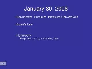
January 30, 2012 • BellRinger: • Complete the logical and spatial puzzles on the handout from Thursday • Objective: • Determine functions and activities to stimulate various parts of the brain • Differentiate between the various stages in cell communication • Homework: • Complete chp 48 notes
Shark Frog Crocodile Cat Human Spinal cord Hind: Medulla oblongata Hind: Cerebellum Optic tectum Bird Midbrain Fore: Cerebrum Olfactory tract Evolution of vertebrate brain forebrain forebraindominant cerebrum hindbrain forebrain
Frontal lobe Parietal lobe Motor cortex Somatosensory cortex Somatosensory association area Speech Frontal association area Taste Reading Speech Hearing Visual association area Smell Auditory association area Vision Temporal lobe Occipital lobe Figure 48.27 • The cerebral cortex controls voluntary movement and cognitive functions • Each side of the cerebral cortex has four lobes • Frontal, parietal, temporal, and occipital
Palm Press Activity • Do not move your feet! • Yoga pose • What area of your brain is active? • Label/color on your brain map
The Cerebellum • Is important for coordination and error checking during motor, perceptual, and cognitive functions • Is also involved in learning and remembering motor skills
Which part of my brain is active? • Attention & alertness? • What else does the part of the brain do? • Label/color on your brain map
The Brainstem • The brainstem consists of three parts • The medulla oblongata, the pons, and the midbrain
Brainstem • The medulla oblongata • Contains centers that control several visceral functions • The pons • Also participates in visceral functions • The midbrain • Contains centers for the receipt and integration of several types of sensory information
The Diencephalon • The embryonic diencephalon develops into three adult brain regions • The epithalamus, thalamus, and hypothalamus
Which part of my brain is active? • Fight or flight? • What controls the breathing/heart rate changes associated with the response? • Why do we have this response – especially to loud noises? • Label/color on your brain map • Listen to someone speak and express themselves in a foreign language • Which part of the brain takes in all sensory input (except smell)
The epithalamus • Includes the pineal gland and the choroid plexus • The thalamus • “Central operator” • Is the main input center for sensory information going to the cerebrum and the main output center for motor information leaving the cerebrum • The hypothalamus regulates • Homeostasis • Basic survival behaviors such as feeding, fighting, fleeing, and reproducing
Emotion Cards • No words, facial expressions only • Which part of your brain is active? • What would happen if we were unable to interpret this nonverbal communication? • Are some people better than others? • Label/color on your brain map
Thalamus Hypothalamus Prefrontal cortex Olfactory bulb Amygdala Hippocampus Figure 48.30 Emotions • The limbic system • Is a ring of structures around the brainstem
This limbic system includes three parts of the cerebral cortex • The amygdala, hippocampus, and olfactory bulb • These structures interact with the neocortex to mediate primary emotions • And attach emotional “feelings” to survival-related functions
Circadian Rhythms • The hypothalamus also regulates circadian rhythms • Such as the sleep/wake cycle • Animals usually have a biological clock • Which is a pair of suprachiasmatic nuclei (SCN) found in the hypothalamus
Short Term Memory • Which part of the brain was stimulated during the memory game? • What part of your brain would be involved if this information was to be stored in your long term memory? • Label/color on your brain map
Eye Input from ears Reticular formation Input from touch, pain, and temperature receptors Figure 48.24 Arousal and Sleep • A diffuse network of neurons called the reticular formation • Is present in the core of the brainstem • Midbrain • Sight/Sound reflexes
A part of the reticular formation, the reticular activating system (RAS) • Regulates sleep and arousal
Midbrain & Hindbrain • How can we see the “ancientness” of these areas of the brain in the functions that these parts control?
Logic Puzzle • What part of the brain was stimulated when you were solving these puzzles? • What is the difference in the way your brain handles detailed, sequential information (logic problem) compared to the way it handles spatial information? • What connects these 2 parts? • Put them together – write your full name in cursive backwards (mirror image) • Label/color on your brain map
Cerebrum • Most highly evolved structure of mammalian brain • Cerebrum divided • hemispheres • left = right side of body • right = left side of body • Corpus callosum • major connection between 2 hemispheres
Lateralization of Brain Function • Left hemisphere • language, math, logic operations, processing of serial sequences of information, visual & auditory details • detailed activities required for motor control • Right hemisphere • pattern recognition, spatial relationships, non-verbal ideation, emotional processing, parallel processing of information
The Cerebral Hemispheres • Cerebral Cortex and Functional Regions • Motor Areas • frontal lobe • What would you do to activate this area? • Which parts of your body do you think have the most motor control? • Sensory Areas • parietal, occipital, and temporal lobes • Think back to the sensory lab…which parts of the body were the most sensitive to touch?
Learning Check • What area of the brain is generally regarded as the area that “makes us human?” What area is so different in our brains?
Central nervous system (CNS) Peripheral nervous system (PNS) Brain Cranial nerves Spinal cord Ganglia outside CNS Spinal nerves Figure 48.19 • In all vertebrates, the nervous system • Shows a high degree of cephalization and distinct CNS and PNS components
Central Nervous System (CNS) • Brain & Spinal cord form the central nervous system • The brain • provides integrative power • Controls complex behavior of vertebrates • The spinal cord • integrates simple responses to certain kinds of stimuli • conveys information to and from the brain
Gray matter White matter Ventricles Figure 48.20 • The brain & spinal cord are hollow • The central canal of the spinal cord and the four ventricles of the brain contain cerebrospinal fluid (CSF)
Peripheral nervous system Somatic nervous system Autonomic nervous system Sympathetic division Parasympathetic division Enteric division Figure 48.21 The Peripheral Nervous System
The PNS transmits information to and from the CNS • The PNS can be divided into two functional components • Somaticnervous system • Carries signals to & from skeletal muscles • Autonomicnervous system • Carries signals to and from internal organs and glands
PNS- Somatic Nervous System • The somatic nervous system • Includes the cranial nerves and spinal nerves • Cranial nerves • originate in the brain • terminate mostly in organs of the head and upper body • Spinal nerves • originate in the spinal cord • extend to parts of the body below the head
PNS • The autonomic nervous system • Regulates the internal environment, in an involuntary manner • Is divided into the sympathetic, parasympathetic, and enteric divisions
Parasympathetic division Sympathetic division Action on target organs: Action on target organs: Dilates pupil of eye Constricts pupil of eye Location of preganglionic neurons: brainstem and sacral segments of spinal cord Location of preganglionic neurons: thoracic and lumbar segments of spinal cord Inhibits salivary gland secretion Stimulates salivary gland secretion Sympathetic ganglia Neurotransmitter released by preganglionic neurons: acetylcholine Constricts bronchi in lungs Relaxes bronchi in lungs Neurotransmitter released by preganglionic neurons: acetylcholine Cervical Accelerates heart Slows heart Inhibits activity of stomach and intestines Thoracic Stimulates activity of stomach and intestines Location of postganglionic neurons: in ganglia close to or within target organs Location of postganglionic neurons: some in ganglia close to target organs; others in a chain of ganglia near spinal cord Inhibits activity of pancreas Stimulates activity of pancreas Stimulates glucose release from liver; inhibits gallbladder Stimulates gallbladder Lumbar Neurotransmitter released by postganglionic neurons: acetylcholine Neurotransmitter released by postganglionic neurons: norepinephrine Stimulates adrenal medulla Promotes emptying of bladder Inhibits emptying of bladder Promotes erection of genitalia Promotes ejaculation and vaginal contractions Sacral Synapse Figure 48.22 • The sympathetic and parasympathetic divisions • Have antagonistic effects on target organs
The sympathetic division • Correlates with the “fight-or-flight” response • The parasympathetic division • Promotes a return to self-maintenance functions- “rest and digest” • The enteric division • Controls the activity of the digestive tract, pancreas, and gallbladder
Summary • Differentiate between the CNS and PNS