Download
1 / 56
560 likes | 769 Views
Chapter 6 The Nervous System The nervous and endocrine systems are the means by which different parts of the body communicate. The nervous system can be separated into the: a- central nervous system, consisting of the brain and spinal cord.

E N D
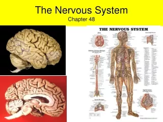
Chapter 6 The Nervous System The nervous and endocrine systems are the means by which different parts of the body communicate. The nervous system can be separated into the: a-central nervous system, consisting of the brain and spinal cord. b-peripheral nervous system, consisting of cranial nerves and spinal nerves that innervate the rest of the body. The coordination of our central and peripheral nervous systems allows us to move, talk, think, and respond.
The Neuron Also called a nerve cell, is the functional unit of the nervous system and is a highly specialized cell. Neural maturation occurs before or soon after birth. Once mature, the neuron does not undergo cellular reproduction and cannot be replaced. Each neuron functions to receive incoming stimuli from, and to send outgoing stimuli to, other nerves, muscles, or glands.
Categories of Neurons - Neurons that carry information from the periphery to the central nervous system are called sensory or afferent neurons. - Neurons that carry information out of the central nervous system to various target organs (muscle cells, other nerves, or glands) are called motor or efferent neurons. - A third group of neurons passes messages between afferent and efferent neurons. These neurons are called interneurons. Almost 99% of all neurons in the body are interneurons, and all interneurons are in the central nervous system. The Synapse A synapse is the point of junction between two neurons. Neurons communicate with each other by releasing chemicals into the small cleft (synaptic cleft) separating one from the other. The chemical released from a particular neuron is called a neurotransmitter.
Neurotransmitters Many neurotransmitters are used in the nervous system. Most neurotransmitters are synthesized in the cell body and transported down the axon to the axon terminal. Because neurotransmitters are released from presynaptic neurons, synaptic transmission usually occurs in one direction: from the presynaptic to the postsynaptic neuron. To respond to a particular neurotransmitter, the postsynaptic cell must have specific receptors for it on its cell membrane.
The Cerebral Cortex The cerebral cortex is the most advanced part of the brain and is responsible for making sense of the environment and initiating thought and goal-oriented behavior. The frontal lobe It contains the motor and premotor areas. Broca's area is in the left frontal lobe and controls the production (or articulation) of speech. The parietal lobe The parietal lobe receives sensory input for touch and pain. The occipital lobe The occipital lobe receives information that originated as signals in the retina of the eye.
The temporal lobe The temporal lobe is the primary association area for auditory information . It is also involved in the interpretation of smell and is important for the formation and storage of memory. The Diencephalon The diencephalon structures lie deep between the cerebral hemispheres. The diencephalon includes the thalamus, the hypothalamus, and the basal ganglia. The thalamus The thalamus receives all incoming sensory information (except smell) . Function of the cerebral cortex depends on thalamic relay.
The hypothalamus It is an important endocrine and neural organ responsible for maintaining homeostasis . The hypothalamus integrates and directs information concerning temperature, hunger, autonomic nervous system activity, and emotional status. It also regulates the levels of several hormones, including the pituitary hormones. The basal ganglia The basal ganglia are important for controlling highly skilled movements that require quickness of response without intentional thought. The Brain stem The brainstem is made of the pons, medulla oblongata, and midbrain. In the brainstem are cells that control cardiovascular and respiratory system functions. Ten of the twelve cranial nerves, controlling motor and sensory function of the eyes, face, tongue and neck, leave from the brainstem.
Reticular Formation Running through the brainstem is a network of many small, branched neurons, called the reticular formation. These neurons include ascending and descending tracts, some of which cluster to form centers that control swallowing, vomiting, and respiratory and cardiovascular reflexes. The reticular formation is also essential for wakefulness and is necessary to focus attention. The Cerebellum The cerebellum helps maintain balance and controls fast, repetitive movements required for activities such as typing, piano playing, and bike riding.
The Spinal Canal(vertebral column) Running down the center of the spinal canal is the spinal cord. It is filled with cerebrospinal fluid (CSF) and surrounded by the bony vertebral column, which extends beyond the end of the spinal tract and offers protection to the delicate nerves inside. Gray and White Matter The spinal cord can be separated into gray and white matter. Gray matter occupies the center of the tract and is filled with interneurons and cell bodies. The white matter, consisting mostly of myelinated ascending and descending tracts.
The Meninges are thin membranes surrounding the brain and spinal cord. There are three meninges: the dura mater on the outside, the arachnoid as a middle layer, and the pia mater lying immediately above the brain. The space above the dura mater is called the epidural, and the space below the dura mater but above the arachnoid is called the subdural. The epidural and subdural spaces contain many small blood vessels. Damage to these vessels leads to blood accumulating in the epidural or subdural spaces. CSF circulates in the subarachnoid space (beneath the arachnoid, above the pia mater).
Cerebrospinal Fluid and the Ventricles Cerebrospinal fluid is a clear fluid surrounding the brain and spinal cord. It is formed as a result of filtration, diffusion, and active transport across special capillaries into the ventricles (cavities) of the brain, especially the lateral ventricle. The capillary network responsible for CSF formation is called the choroid plexus. The Blood-Brain Barrier The blood-brain barrier results from tightly fused endothelial cells present in the brain capillaries that limit diffusion and filtration. The blood-brain barrier protects delicate brain cells from exposure to potentially harmful substances. Many drugs and chemicals cannot cross the blood-brain barrier.
Brain Blood Flow and Brain Metabolism The brain receives approximately 15% of the cardiac output. This high rate of blood flow is required to meet the brain's continually high demands for glucose and oxygen. The brain is unique in that it normally uses only glucose for the production of ATP. Unlike other cells, brain cells do not store glucose as glycogen; therefore, the brain must continually receive oxygen and glucose through brain blood flow. Oxygen deprivation for as little as 5 minutes, or glucose deprivation for 15 minutes, can cause significant brain damage.
Intracranial Pressure The pressure inside the cranium is called intracranial pressure (ICP). ICP is determined by: - the volume of blood in the brain - the volume of CSF - and the volume of brain tissue. Normally, ICP ranges from 5 to 15 millimeters of mercury (mmHg). The Peripheral Nervous System Consists of 12 spinal nerve pairs and 31 cranial nerve pairs . In all spinal nerves and most cranial nerves, afferent and efferent fibers travel together in opposite directions. Some cranial nerves carry only afferent information(pure sensory). Afferent neurons convey information to the central nervous system. Efferent neurons deliver neural stimulation to muscles and glands.
The Autonomic Nervous System Autonomic nerve fibers leave the spinal cord and innervate smooth and cardiac muscle and the endocrine and exocrine glands. Autonomic nerves fibers are considered involuntary because there is little conscious control over their function. The two divisions of the autonomic nervous system, the sympathetic and parasympathetic divisions. Sympathetic and parasympathetic nerves innervate many of the same organs but typically cause opposite responses. The cell bodies of these neurons lie in the brain or spinal cord.
Pathophysiologic Concepts Brain Death Brain death is irreversible loss of cerebral hemisphere, brainstem, and cerebellum function. Consciousness is lost, as is maintenance of respiration, cardiovascular, and temperature control function. There is no sleep-wake cycle, no pain response, and no reflexes . The electroencephalogram (EEG) is flat. Dementia Dementia is a loss of intellectual functioning without a loss of arousal functioning. Memory, general knowledge, judgment, and interpretation of written and oral communication may be affected. Dementia may be caused by infection, drugs, trauma, or tumors.
Increased Intracranial Pressure It increases with increases in cranial blood, CSF, or tissue. Intracranial hypertension causes delicate neurons and capillaries in the brain to become compressed, leading to hypoxia, neuronal injury and death . If intracranial pressure reaches systemic mean arterial pressure, blood flow to the brain will stop and the individual will die. Causes of Increased Intracranial Pressure • - Anything that significantly increases CSF production or blocks CSF outflow . • - Any increase in tissue mass (e.g., that associated with a growing brain tumor) • - Edemadue to infection and inflammation • - Severe trauma to the head, a burst aneurysm, or a hemorrhage in the brain,
The Stages of Intracranial Hypertension The response of the brain to increased intracranial pressure is called compensation. The brain goes through four stages : Stage 1 An increase in one of the three volumes in the brain (blood, CSF, or tissue) is normally compensated for by a decrease in one or both of the other volumes. Usually, this stage involves decreased CSF production or increased CSF reabsorption, followed by increased arterial constriction to decrease blood flow into the brain.. Stage 2 The brain responds by constricting cerebral arteries reducing blood flow, so may leads to cerebral hypoxia and hypercapnia and deterioration of brain function. Clinical signs include decreased level of consciousness, alterations in breathing pattern, and pupillary changes.
Stage 3 In response to worsening hypoxia and hypercapnia, the cerebral arteries undergo reflex dilation, with the goal of increasing brain oxygen delivery. As blood volume increases, however, intracranial pressure increases further, thereby worsening the situation. This cycle of increasing hypoxia leading to increasing pressure, thereby worsening the hypoxia, is called decompensation. The result is a pronounced decrease in consciousness, altered respiratory pattern, and loss of pupillary reflexes. Stage 4 As the swelling and pressure in one compartment of the brain become very high, herniation (bulging) into another compartment occurs. When intracranial pressure reaches mean systolic pressure, cerebral perfusion stops.
Treatment of Intracranial Hypertension • Effective monitoring of intracranial pressure (ICP). • Osmotic diuretics (mannitol) to reduce blood volume • Steroids to decrease inflammation. Tests of Neurologic Functioning - Electromyography(EMG) Measures peripheral nerve function and is used to diagnose, describe, and monitor neuromuscular pathology - Electroencephalography (EEG) Measures electrical activity occurring in the brain through electrodes placed on the scalp. This technique offers a fast, picture of brain activity.
- Magnetic Resonance Imaging (MRI) Captures what is happening in the brain physiologically before, during, and after an individual performs a task. - Computed Tomography (CT) Involves computer analysis of multiple radiologic images. In a CT scan, an x-ray beam is rotated around the patient, and passes successively through tissue from multiple directions. - Positron Emission Tomography(PET) Involves intravenous injection or inhalation of a positron-emitting isotope, followed by sequential radiographs of the skull that monitor the decay of the isotope in tissues that take up the label. - Ultrasonography Use reflected sound to measure blood flow velocity, which is important for assessment of cerebral blood flow when evaluating ischemic cerebrovascular disease.
Conditions of Disease or Injury 1-Seizure Disorder Seizures result when certain cerebral neurons exist in a hyperexcitable or easily depolarized state. Neurons of the epileptogenic focus respond to levels of stimuli that do not produce discharge in other neurons. If the seizure arises from a discrete focus and is limited to one side of the brain, it is called a partial seizure, and consciousness is usually not lost. Partial seizures may progress and become generalized. The time of unconsciousness after any generalized seizure is called the postictal phase. **If one seizure is followed by a second or third seizure before the individual regains consciousness, status epilepticus is said to occur.
Seizure Syndromes - Generalized seizures characterized by sudden onset of rigid, intense contractions of arm and leg muscles (tonic seizure), followed by rhythmic contraction and relaxation of the muscles (clonic seizure). Generalized seizures may occur idiopathically (for no known reason) or after brain trauma, infection, tumor, or bleeding. - Partial or focal seizures are simple partial seizures, during which consciousness is not impaired .It may occur idiopathically or after brain damage. Consequences of a Seizure - During a seizure, cerebral oxygen demand increases more than 200%. If this oxygen demand cannot be met, brain hypoxia and brain damage may occur. - Seizures that continue for extended periods, or the occurrence of status epilepticus, greatly increase the chance of brain damage. - Social isolation and reduced employment.
Causes of Seizure Disorders: • severe hypoxemia (decreased oxygen in the blood), • hypoglycemia (decreased glucose in the blood), • acidemia (increased acid in the blood), • alkalemia (decreased acid in the blood), • dehydration, water intoxication, • high fever. • Drug withdrawal or abuse, and toxemia of pregnancy. N.B. Syncope (fainting) is often misdiagnosed as a seizure because some muscle movements may be similar. Unconsciousness and muscle jerking related to fainting rarely last longer than 5 to 10 seconds, and fainting is not associated with postictal symptoms such as fatigue.
2- Epilepsy Epilepsy refers to a seizure that occurs without a reversible, metabolic cause. Epilepsy may be: • Primary epilepsy develops spontaneously, usually in childhood, and has a genetic predisposition • Secondary epilepsy occurs as a result of hypoxemia, head injury, infection, stroke, or central nervous system tumor. Clinical Manifestations *Partial seizures may be associated with: - Facial movements or grimaces - Jerking beginning in one part of the body, which may spread. - Sensory experiences of sights, smells, or sounds.
- Tingling - An alteration in level of consciousness *Generalized seizures may be associated with: - Unconsciousness. - Uncontrolled jerking of arms and legs - A short period of apnea (breathing cessation) - Salivation and frothing at the mouth - Tongue biting - Incontinence - A postictal stage of stupor or coma, followed by confusion, headache, and fatigue. - A prodroma may occur with any seizure type. A prodoma is a certain feeling or symptom that may precede a seizure by hours or days. - An aura may occur with any seizure type. An aura is a certain sensory sensation that frequently or always immediately precedes a seizure.
Diagnostic Tools - Basic laboratory evaluation must be performed to rule out metabolic causes or drug-induced seizures. - Lumbar puncture is performed to rule out meningitis or encephalitis . - MRI to identify brain lesions such as tumor, abscess, or vascular malformation. - A CT scan. - An EEG may allow diagnosis of the type and location of the seizure. Complications - Hypoxic brain damage. - Depression and anxiety. Treatment - The goal of seizure treatment is zero seizure occurrence with a minimum of treatment-induced side effects. - The medication selected must be appropriate for the seizure type. - Resective surgery to excise the epileptogenic focus is becoming more common
3-Head Injury Head injuries may be open (penetrating through the dura mater) or closed . Types of Head Injuries a-Concussion A concussion is a closed head injury usually characterized by loss of consciousness. Concussion results in a brief period of apnea. A concussion can be mild, moderate, or severe, depending on the length of time the person is unconscious. b-Epidural Hematoma An epidural hematoma is the accumulation of blood above the dura mater. An epidural hematoma occurs acutely and is usually caused by a life-threatening arterial bleed. c-Subdural Hematoma A subdural hematoma is the accumulation of blood under the dura mater, but above the arachnoidmembrane.A subdural hematoma may develop rapidly(acute ) or may result from a slow bleed, in which case it is called a subacute subdural hematoma.
d-Subarachnoid Hemorrhage Is the accumulation of blood under the arachnoid membrane, but above the pia mater. This space normally contains only CSF. It usually results from a burst intracranial aneurysm, severe hypertension, an arteriovenous malformation, or a head injury. Clinical Manifestations - With a concussion, consciousness is often lost. - Respiratory patterns may become progressively abnormal. - Pupillary responses may be absent . - Headache. - Vomiting may occur as a result of increased intracranial pressure. - Behavioral, and physical changes in speech and motor movements may occur immediately or develop slowly. - Amnesia related to the event is common.
Diagnostic Tools - A skull radiograph may locate fractures or a developing bleed or blood clot. - A CT scan or an MRI may pinpoint the site and extent of injury. Treatment - Mild and moderate concussions are usually treated with observation and bed rest. - Surgical ligation of a bleeding vessel and evacuation of a hematoma may be required. - Surgical debridement (removal of foreign material and dead cells) may be required, - Decompression through the drilling of holes into the brain. - Mechanical ventilation may be required. - Antibiotics are required for open head injury to prevent infection. - Methods to decrease intracranial pressure .
5-Spinal Shock Involves immediate loss of all spinal reflexes. Lost reflexes include those controlling posture, bladder and bowel function, blood pressure, and maintenance of body temperature. Spinal shock typically lasts 7 to 21 days, but may last longer. 6-Paralysis Paralysis is the loss of sensory and voluntary motor function. With spinal cord transection, paralysis is permanent. - Paralysis of the upper and lower extremities occurs with transection of the cord at level C6 or higher and is called quadriplegia. - Paralysis of the lower half of the body occurs with transection of the cord below C6 and is called paraplegia. If only one half of the cord is transected, hemiparalysis may occur.
Clinical Manifestations • - Loss of sensation, motor control, and reflexes below the level of injury. Diagnostic Tools • Physical examination coupled with CT and MRI will document vertebral and spinal injury and edema. Complications If damage and swelling around the cord is in the cervical spine (down to approximately C5), respirations may cease because of compression of the phrenic nerve, which exits between C3 and C5 and controls the movement of the diaphragm.
Treatment - Immobilization to prevent cord severing or additional damage - Early surgical intervention to relieve pressure on the cord - Immediate (within the first hour) large-dose administration of steroids has been shown to reduce cord swelling and inflammation and limit the extent of permanent damage. - Surgical fixation of the vertebral column hastens and supports healing. - Physical therapy is begun soon after the patient's condition stabilizes.
7-Cerebral Vascular Accident (CVA) Often called a stroke or a brain attack, is a brain injury related to an obstruction in brain blood flow. There are two types of CVAs: ischemic and hemorrhagic. Ischemic Stroke :Result of a thrombus or an embolus. A thrombotic stroke usually due to severe atherosclerosis. Frequently, an individual will experience one or more transient ischemic attacks (TIAs) before a true thrombotic stroke occurs. A thrombotic stroke typically develops over a period of 24 hours. During the period in which a stroke is progressing, the individual is said to be suffering from a stroke in evolution. At the end of that period, the individual is said to have suffered a completed stroke. An embolic stroke develops often after arterial occlusion by an embolus formed in the heart after a myocardial infarct or atrial fibrillation.
Hemorrhagic Stroke :occurs when a blood vessel in the brain is broken by burst aneurysm, or an arteriovenous malformation (abnormal connection). Hemorrhage into the brain significantly increases intracranial pressure, worsening the resulting brain injury. Clinical Manifestations - Symptoms of a TIA may include temporary numbness of the face or limbs, slurring of words, confusion, dizziness, and changes in vision. If any of these occurs, an individual should immediately seek medical assistance. - With a CVA, the area of the brain that becomes ischemic determines the presenting clinical manifestations. Mentation, emotions, speech, vision, or movement can be affected. Many changes are irreversible, but some are reversible. - A hemorrhagic stroke is frequently accompanied by a severe headache and loss of consciousness.
Diagnostic Tools - Rapid diagnosis of a CVA is essential to minimize damage. - CT scan is the method of choice for assessment of an acute presentation of a CVA. - MRI devices are used less often in this emergency situation. Complications - A major CVA to the part of the brain controlling respiration or cardiovascular response may lead to death - Communication difficulties. Hypoxia of motor areas in the brain may lead to - paresis. - Emotional changes .
Treatment - If ischemic in nature, thrombolytic agents, such as tissue plasminogen activator (TPA), can be administered. TPA should be given as early as possible (at least within the first 3 hours of the attack) - A hemorrhagic stroke is treated with emphasis on stopping the bleeding and preventing another occurrence. Surgery may be required. - All stroke patients are treated with bed rest and a reduction of external stimuli to reduce cerebral oxygen demands. - Measures to reduce intracranial edema and pressure may be instituted. - Physical, speech, and occupational therapy are often required.
8-Central Nervous System Infection central nervous system infection may involve the brain tissue (encephalitis), or the meninges (meningitis). • A-Encephalitis • Encephalitis is usually a viral infection of the brain. It is often carried by a mosquito vector or related to infection with herpes simplex 1 or cytomegalovirus. Nerve cell degeneration is widespread and edema and swelling are severe.
b-Meningitis Meningitis is the most common serious infection of the CNS. It is usually caused by bacteria or a virus, although fungi, protozoa, and toxins are also causes. Meningitis frequently occurs from the spread of an infection elsewhere in the body, for example, the sinuses, ears, or upper respiratory tract. A posterior basilar skull fracture with a ruptured eardrum may also cause meningitis. Secondary encephalitis may occur. Even when treated, up to 40% of meningitis cases are fatal and up to 30% of survivors have neurological complications.
Meningitis now occurs most commonly in adults ages 19 to 59. In this age group, the most common cause of bacterial meningitis is Streptococcus pneumoniae (pneumococcal meningitis). The next greatest incidence is in children ages 2 to 18, and the cause is most often Neisseria meningitides (meningococcal meningitis). In the neonate, the cause is most often group B streptococcus. In infants aged 1 to 23 months the causes are split almost equally between S. pneumoniae and N. meningitidis.
Clinical Manifestations • Symptoms of increased intracranial pressure including headache, decreased consciousness, and vomiting. • Papilledema (swelling of the area around the optic nerve) may occur in severe cases. Typically, the symptoms are worse with encephalitis. • Fever from infection is common . • Photophobia (painful response to light). • Inability to flex the chin to the chest without pain (nuchal rigidity). • Encephalitis typically presents with dramatic signs of delirium and a progressive decrease in consciousness. • Seizures and abnormal movements may occur.
Diagnostic Tools • - CSF with elevated protein, elevated neutrophils, and low glucose in the CSF indicate meningitis. • - CT scan and MRI may be used to evaluate the degree of swelling and sites of necrosis. CT is very rapid and is most useful in emergency situations. Complications • Permanent disability,brain damage, or death from encephalitis or, less commonly, meningitis. • Seizures may develop. Treatment • - A broad spectrum antibiotic after CSF culture results. • - An antiviral drug will be administered for encephalitis. • - Measures to reduce intracranial pressure will be initiated, especially for encephalitis. • - Some types of meningitis will require the patient to be isolated in the hospital.
10-Alzheimer Disease Alzheimer disease is a progressive dementia characterized by the widespread death of brain neurons. Alzheimer disease typically develops after the age of 65, causing senile dementia. However, it may occur earlier and result in presenile dementia. There is a genetic tendency to develop the disease. Clinical Manifestations Insidious, slowly progressing forgetfulness, decreased judgment, behavioral and personality changes developing over a period of up to 10 years.
Diagnostic Tools - Eliminating metabolic or vascular causes of the mental deterioration, approximately one-third of suspected dementia cases are caused by reversible disorders, including metabolic imbalance, drug effects, CVA, vitamin deficiencies, and depression. These causes must be ruled out using CT or MRI, a complete blood count, and metabolic studies. Treatment -Patient and family education regarding memory aids, diet, and safety issues may slow the progression of symptoms. - Medications (Cognex) for the slowing or reversal of early Alzheimer symptoms are available and may delay the progression of symptoms in some patients.
11-Parkinson Disease • Is a progressive brain disorder, typically occurs in the sixth or seventh decade of life. • Cause • -The cause is unknown. There is at least some genetic tendency toward developing the disease. • -Viruses and toxins have been implicated in some studies. • - sometimes ,brain trauma, infection, or tumors. • Clinical Manifestations • - A tremor at rest. • - Drooling and dysphagia (difficult swallowing). • - Muscle rigidity, and stiffness.
Diagnostic Tools • history and physical examination. • A positive response to levodopa is strongly indicative of parkinson. Complications • Many patients with Parkinson disease develop dementia. Treatment • - Dopaminergic (L-dopa) or anticholinergic drugs may be administered to reduce symptoms. • -Transplanting cells from the basal ganglia or adrenal medulla (where dopamine is also produced) of fetuses into the brains of patients with Parkinson disease has been successful in some studies.
12-Myasthenia Gravis • Is a peripheral nervous system disorder characterized by autoantibody production against the receptors for acetylcholine present on the motor end-plate region of skeletal muscles. • It may first cause weakness of the muscles controlling eye movements (ocular myasthenia gravis) or may affect the entire body (generalized myasthenia gravis). • The cause of myasthenia gravis is unknown but appears to be associated with a familial tendency toward developing autoimmune disease.
Clinical Manifestations • - Weakness of the muscles of the eyes, causing ptosis (drooping of the eyelids). • - Weakness of face, neck, and throat muscles, causing difficulty eating and swallowing. • - Continued spread of muscle weakness. • Initially there is easy fatigue with recovery of strength after rest. Eventually there is no recovery of strength after rest. Complications • - Myasthenic crisis, characterized by severe worsening of skeletal muscle function culminating in respiratory distress and death as the diaphragm and intercostal muscles become paralyzed, may occur after a stressful experience such as an illness, emotional upset, surgery, or during pregnancy. • - Respiratory distress may occur.
Treatment • - Frequent rest periods during the day conserve strength. • - Anticholinesterase medications are provided to prolong the half-life of acetylcholine at the neuromuscular junction. 13-Guillain-Barr Syndrome It results from an autoimmune attack against the myelin surrounding the peripheral nerves. With destruction of the myelin, the axons can be damaged. The disease usually occurs 1 to 4 weeks after a viral nfection or immunization. The muscles of the lower extremities are usually affected first, with paralysis advancing up the body. Respiratory muscles may be affected, leading to respiratory collapse. Clinical Manifestations Ascending muscle weakness or paralysis.
Diagnostic Tools • Nerve conduction tests will demonstrate neuronal dysfunction. • Elevated protein in the CSF is common. Complications • Respiratory or cardiovascular collapse may cause death. • Weakness of some muscles may persist. Treatment • - Ventilator support may be required if the respiratory muscles are affected. • - Anti-inflammatory medications may limit the autoimmune attack.
14-Spina Bifida Spina bifida is a congenital neural tube defect characterized by a failure of the vertebral arches to close. This results in a cyst-like protrusion of the meninges alone (meningocele) or of the meninges and the spinal cord (myelomeningocele) out of the vertebral column. Causes of Spina Bifida Although the cause of spina bifida is unknown, a genetic predisposition may exist. Increased risk of the disorder occurs with maternal folic acid deficiency. Folic acid deficiency is common in women; therefore, it is strongly recommended that all women anticipating pregnancy begin taking folic acid vitamin supplements at least 3 months before conception.