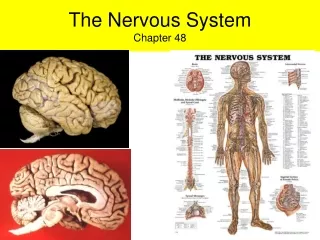
The Nervous System: Functions, Structure, and Impulses
370 likes | 397 Views
Explore the complexities of the nervous system, including its coordination with the endocrine system and the differences between the central and peripheral nervous systems. Learn about neuron structure, action potentials, and the significance of synapses in nerve impulse transmission. Understand how neurons distinguish between strong and weak stimuli and the importance of myelin sheaths in the nervous system.

The Nervous System: Functions, Structure, and Impulses
E N D
Presentation Transcript
Although the Nervous System and the Endocrine System coordinate responses, there are 3 main differences. The nervous system: is much faster has pinpoint control has more structural complexity
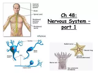
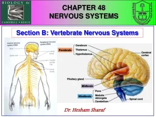
Nervous system = 2 main parts: Central Nervous System (CNS) = brain & spinal cord Peripheral Nervous System (PNS) = nerves (throughout body) including: Afferent nerves: sensory Efferent nerves: motor
Efferent PNS has 2 main divisions Somatic PNS: responds to external environment (mostly voluntary) Ex. Skeletal muscle Autonomic PNS: regulate internal env (mostly involuntary) Ex. Invol. Muscle or glands
Autonomic PNS is further subdivided into: Sympathetic: stimulates organs & energy use (for action) Parasympathetic: causes energy intake and conservation – antagonistic
3 Overlapping functions of the Nervous System: sensory input: afferent neuron integration: info is interpreted and associated w/ responses by CNS motor output: efferent neuron sends signal to effector cells (muscle or gland)
Neuron Structure: Dendrites: short, highly branched fibers. Receive stimuli and send electrical impulses to cell body. cytoplasm, nucleus, most organelles Cell Body: Axon: long, branched extension of cell body – conducts nerve impulse to another neuron or effector Axon (synaptic) terminals: release neurotransmitters Synapse = gap between synaptic node and next neuron or effector
Nerve impulse: Travels in one direction ONLY! Receptor cell body dendrite axon synapse
PNS Axon Structure: Most of the axons in the PNS have 2 coverings made of supportive glia cells : Both called Schwann cells Cellular sheath: aids in nerve regeneration Myelin (inner) sheath: membrane contains myelin which provides electrical insulation Nodes of Ranvier: gaps in myelin sheath – axon not insulated (allows impulse to jump)
Multiple Sclerosis: neurological disease – deterioration of myelin sheath Causes disruptions in nerve impulse transmission, leads to – loss of coordination &/or paralysis
CNS Axon Structure: The axons in the CNS are supported and insulated by glia cells Two types: Oligodendrocytes: insulate axons (w/ myelin) Astrocytes: encircle capillaries to form a blood-brain barrier
Note: Not all neurons are exactly alike. We’re focusing on a typical PNS neuron. When a PNS neuron is wrapped in Schwann cells (like we discussed), the axons are called “white fibers.” (“white matter” in the brain is made of glial cells & myelinated axons) If, instead, several axons are held together by a single Schwann cell that does NOT wrap around them, the axons are called “gray fibers.” (“grey matter” in brain is cell bodies and is unmyelinated)
Nerve = Bundle of hundreds of axons wrapped together in connective tissue.
Action Potentials in Neurons Do you Remember any of this??? All cells have a resting membrane potential: net difference in charge across plasma membrane Inside of cells is negatively charged due to: large neg. charged organic molecules trapped inside, Limited permeability to + ions, and the action of the Na+/K+ pump Channel Proteins (in membrane): “gates” for Na+ or K+, voltage regulated
In Neurons An or disturbance near the dendrite causes a local which opens the Na+ gates, and Na+ rushes ______. electrical chemical depolarization in The depolarization causes an opening of the K+ gates and -- K+ moves out to cause repolarization The flow of ions and resulting change in membrane potential = action potential
All-or-None Law: once depolarization starts, the entire sequence occurs (regardless of stimulus size) How, then does the nervous system distinguish between strong and weak stimuli? Strong Stimuli = higher frequency of action potentials (neuron “fires” repeatedly)
Depolarization Speed is increased with: thicker axons Saltatoryconduction: myelin sheath insulation prevents movement of ions through membrane. Therefore, action potentials only occur at gaps (nodes of Ranvier) which are spaced about 1mm apart. Action potential: leaps from node to node.
sends to the through electrical or chemical synapses. Presynaptic cell postsynaptic cell ElectricalSynapses: Cells must be connected by – gap junctions to allow ion currents to flow Responsible for certain rapid movements in some vertebrates (CNS): escape from predator… Much less common than chemical synapses in vertebrates (& many invertebrates)
ChemicalSynapses: Cells are not connected: action potential cannot cross synapse Electrical signal of presynaptic cell must be converted to a signal which crosses the synapse and is converted back to an electrical signal in the postsynaptic cell. chemical Synaptic terminal (axon end): contains many synapticvesicles full of neurotransmitters When the action potential reaches the terminal, it causes Ca+ to rush into cell through voltage-sensitive channels. The sudden influx of Ca+ causes synaptic vesicles to fuse with the presynaptic membrane and release neurotransmitters via: exocytosis.
Neurotransmitters binding to postsynaptic membrane: alter the membrane potential Example: binds to gate channels in postsynaptic membrane to allow Na+ & K+ diffusion. The enzyme, cholinesterase, breaks it down for . (restores resting potential) Acetylcholine recycling
Neurotransmitters Can excite membranes by: bringing the voltage closer to the threshold potential Can inhibit membranes by: hyperpolarizing the membrane
Excitatory Postsynaptic Potentials (EPSP’s) Neurotransmitter opens Na+ & K+ gates – generates action potential Summation: cumulative effect of postsynaptic potentials. 2 types: temporal: chemical transmissions occurring in quick succession (no time to return to resting potential) spatial: several different presynaptic neurons have an additive effect on the postsynaptic neuron AT THE SAME TIME
Inhibitory Postsynaptic Potentials (IPSP’s) Neurotransmitter causes K+ to rush out (or Cl- to enter) – hyperpolarizes the membrane Makes it much harder to generate action potential dopamine, Example Neurotransmitters: norepinephrin, serotonin, glutamic acid…
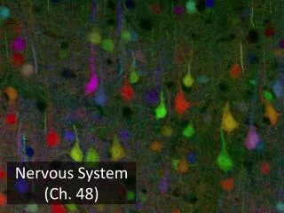
Invertebrate Nervous Systems Many invertebrates have ___ ventral ___ nerve cords and segmental ganglia: Clusters of nerve cell bodies. Invertebrate neurons: A. A dye-filled antennal neuron, within a Drosophila ganglion B. Giant neuron in a cricket ganglion C Leech ganglion from an embryo (with fluorescent dyes) D. Two auditory neurons in the crickets ganglion Source: http://www.scholarpedia.org/article/Neuron
Vertebrate Nervous Systems Vertebrates have ___ dorsal ___ nerve cords without segmental ganglia: Segmented ganglia lie just outside the cord. Invertebrate neurons: A. A dye-filled antennal neuron, within a Drosophila ganglion B. Giant neuron in a cricket ganglion C Leech ganglion from an embryo (with fluorescent dyes) D. Two auditory neurons in the crickets ganglion Source: http://www.scholarpedia.org/article/Neuron
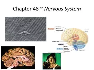
Embryonic Development of the Brain All vertebrates have three bilaterally symmetrical anterior bulges of the neural tube: Forebrain, midbrain, hindbrain During vertebrate evolution, the brain divided further. Birds and mammals have a much larger: forebrain See Figure 48.23, p. 1028
The Brainstem • “Lower brain” • 3 parts: medulla oblongata, pons, midbrain • Structure: stalk at anterior end of spinal cord with caplike swellings
The Brainstem: medulla and pons • Functions: • Release neurotransmitters which cause changes in: attention, alertness, appetite, and motivation • Control visceral (automatic, homeostatic) functions such as: breathing, heart / vessel activity, swallowing, vomiting and digestion
The Brainstem: medulla and pons • Controls information transmission: input from sensory neurons and motor instruction output travel through brainstem • Coordinates large-scale body movements --most axons carrying info. between spinal cord and mid- or forebrain cross over here: causing left side of brain to control movements on right side (and vice versa)
The Brainstem: midbrain • Contains centers for receipt and integration of sensory information: hearing (both) and • vision (receipt & reflexes) The Brainstem: reticular formation Neuron network throughout the brainstem which filters sensory input to regulate: Sleep and arousal
The Cerebellum • Functions: • ___ Coordination ___ during motor, perceptual and cognitive functions. • Learning and remembering ___ motor ___ skills such as bike riding. • Coordinates movements and balance such as in • hand eye coordination.
The Diencephalon Found in Embryo: Develops into epithalamus, thalamus and hypothalamus. • Epithalamus includes: pineal gland & choroid plexus (capillary cluster) • Thalamus: processes input / output of info between senses, cerebrum & motor neurons • Hypothalamus: homeostatic regulation via hormones, basic survival mechanisms, mating behaviors, and pleasure
The Cerebrum • Structure: • Divided into left and right ___ hemispheres___. • Each side has: gray matter -- (outer covering) cerebral cortex • internal white matter • basal nuclei – (groups of • neurons involved in movement)
The Cerebrum: cerebral cortex • Largest and most complex part of human brain. • Functions: sensory info is analyzed, motor commands are issued, language is generated • Neocortex (outermost part of mammalian cerebrum) is highly convoluted in humans to provide: Huge surface area and about 80% of brain’s total mass.
The Cerebrum: corpus callosum • Thick band of ___ axons ___. • Function: enables communication between right and left cerebral cortex • Damage to one area of the cerebrum early in development can cause redirection of its functions to other areas. Even in adults, cerebral cortex damage can lead to: new brain circuits.