
Local Skin Flaps
E N D
Presentation Transcript
1. Local Skin Flaps Kevin Katzenmeyer, MD
Karen Calhoun, MD
June 21, 2000
2. Introduction Facial defects common
Trauma
Skin malignancies
Treatment
secondary healing
skin graft
local flaps
3. Paradigm Etiology
Cosmesis
Function
Patient�s wishes
Surgeon�s experience
4. Local Flaps - Classification Blood supply
random
axial
Tissue movement
rotation
advancement
transposition
5. Local Flaps - Blood supply Segmental arteries
Perforating branches
Direct cutaneous vessels
Subdermal plexus
6. Random flaps Most common
Based on subdermal plexus
Unpredictable
Length:width of 3:1 or 4:1
7. Axial flaps Limited by available vessels
Based on direct cutaneous vessels
Random flap at distal tip
Examples
nasolabial
midline forehead flaps
8. Flap survival Length:Width
increased width of
base would increase
surviving length but
feeding vessels have
same perfusion pressure
Perfusion pressure
9. Blood supply Supply exceeds requirements
Changes
temperature
autonomics
trauma
Arteriovenous shunts
sympathetic control
fully opened shunt bypasses capillary bed
10. Arteriovenous Shunts
11. Delay phenomenon Incise and undermine
10 to 21 day delay most common
No benefit at 3 wks to 3 mos
Improved blood supply
AV shunt closure
conditioning to ischemia
alignment of vessels
12. Skin stretch Elasticity
elastin
collagen
tension vs. blood supply
13. Skin biomechanics Creep
extrusion of fluid in dermis
breakdown of dermal framework
Stress relaxation
increased cellularity
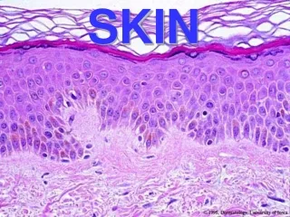
14. Skin characteristics RSTLs
LMEs
15. Facial Aesthetic Units
16. Planning History
PVD/CAD, collagen vascular dz, DM, XRT
Social habits
cigarettes?
Medications
ASA, NSAIDs, anticoagulants
Cause of defect
recurrence?
17. Physical Exam Defect
size, placement
Surrounding skin
lesions, laxity, color match, scars
Facial structures
functional concerns, lip, lid
Incision placement
RSTLs, BAUs
18. Planning Template
Draw options/Measure
Undermine
Review options/Remeasure
Incise
Rotate vs. advance vs. transpose
Key stitches
Excise cones
Close
19. Flap types Rotation
Advancement
Transposition
Not concrete, variations exist
20. Rotation Pivotal flap
curvilinear
standing cone results
two borders
broad based
Uses - cheek, forehead
21. Rotation flap
22. Transposition Rhomboid, dufourmental, bilobed
Linear axis
Rotated over intact skin
Pivot point
Versatile
23. Transposition Geometry
measure, remeasure
Rhomboid
60 & 120 degree angles
Dufourmental
60 to 90 degree angles
4 choices
24. Rhomboid flap
25. Dufourmental Flap
26. Bilobed Double transposition flaps
Original description
90 degree arcs
final 180 degree arc
Arcs of 90 to 110 degrees preferrable
Uses - lower third of nose
27. Bilobed Flap
28. Advancement Sliding movement
adequate undermining
standing cones created
Types
monopedicle, bipedicle, V-Y, A-T, cheek
Uses - forehead, brow
29. Monopedicle Forehead, Brow
3:1 ratio
Burow�s triangles
30. Bipedicle Forehead, Brow
Disadvantage
long suture line
31. V-Y flap
32. A-T flap Bilateral advancement
triangular defect
Uses - hairline, brow, lip
33. Cheek advancement Advancement
Some rotation
Uses - medial cheek, nasofacial sulcus
Prevent complications (ectropion)
34. Cheek Advancement Flap
35. Nasolabial flap Axial pattern - angular artery
Inferior and superior flaps
Uses - lower 2/3 of nose, perinasal area, upper lip
pin cushioning, blunting of nasofacial sulcus
potential ectropion, scleral show
36. Nasolabial Flap Inferiorly based
37. Nasolabial Flap Superiorly based
38. Midforehead flap Indian rhinoplasty
Median, paramedian forehead flap
axial pattern
supratrochlear artery - at medial brow, 2 cm from midline
pedicle can be as little as 1.2 cm
thin distal tip appropriately
Disadvantages
long scar, limited length, revision
39. Midforehead Flap
40. Paramedian Forehead Flap
41. Paramedian Forehead Flap
42. Postoperative Care Pain reliever
Wound care
hydrogen peroxide, antibiotic ointment
Sutures removed at 5-7 days
Direct sunlight avoided for 2-3 months
Dermabrasion - 6-12 weeks
Revision/Irregularization - 6 months
43. Complications Infection
Hematoma/ seroma
Cyanosis
Failure/necrosis
44. Case Presentations
Pick the flap
45. Example #1 Left temple defect
46. Intraop
47. Example #2 Chin defect
48. Outcome