Download
1 / 29
290 likes | 1.3k Views
Tensor Fascia Lata , Rectus Femoris , Gracilis Flaps. Ian Maxwell. Tensor Fascia Lata. Overview. Classification: Mathes and Nahai type I muscle flap Pedicle length: up to 10cm Pedicle calibre : 1.5-2.5mm (artery) Uses Groin Ischium Perineum Lower abdomen Sacrum Trochanter.

E N D
Tensor Fascia Lata, Rectus Femoris, Gracilis Flaps Ian Maxwell
Overview • Classification: Mathes and Nahai type I muscle flap • Pedicle length: up to 10cm • Pedicle calibre: 1.5-2.5mm (artery) • Uses • Groin • Ischium • Perineum • Lower abdomen • Sacrum • Trochanter
Anatomy • Origin: • anterior iliac crest • Insertion: • illiotibial tract • Blood supply: • ascending branch of the lateral circumflex femoral artery • Innervation: • Sensory: lateral cutaneous sensory nerve of the thigh • Motor: distal branch of the superior gluteal nerve
Variations • Bone (ASIS) +/- TFL muscle • Neurosensory • Include lateral sensory branch of T12 or lateral femoral cutaneous nerve • Expanded or transverse skin paddle • Perforator flap • V-Y advancement
Surgical Markings • Mark a line from ASIS to the lateral patella (anterior axis) • Pedicle is located anywhere from 8-12 cm below ASIS • Axis of femur marks posterior aspect of flap
Dissection • Elevate flap from distal to proximal • More proximally, retractor placed between rectus femoris and vastuslateralis • First identify descending branch of LFCA, then identify ascending branch more superiorly • Isolate pedicle course to TFL • Pedicle can be traced to lateral femoral circumflex vessels/profundus to gain calibre and length
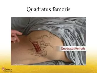
Overview • Reliable blood supply and motor innervation • Classification: Mathes and Nahai type 2 (dominant and minor arterial supply) • Major disadvantages are: • Not a particularly expendable muscle • Pedicle can be affected by atherosclerosis • Coverage of lower abdomen, groin, ischium, trochanteric region, functional muscle transplantation
Anatomy • Origin: iliac portion of acetabulum and ASIS • Insertion: patella • Arterial supply: • Dominant = descending branch of lateral femoral circumflex artery • Minor = ascending branch of lateral femoral circumflex artery and muscular branch of SFA • Vein = venae commitantes • Pedicle length/calibre= 5cm/2mm • Nerve = motor branch from femoral nerve
Variations • With or without skin paddle • Most perforators within middle third of thigh • Innervated functional muscle
Surgical Markings • Draw line from ASIS to mid patella for longitudinal axis • Lazy Sincision over muscle • Pedicle just proximal to junction of proximal and middle thirds of thigh (8-10 cm below AIIS) • Length limited to middle 1/3rd of thigh
Flap dissection • Incise through skin paddle (if required) • Through muscular fascia • Sartorius and rectus femoris identified • Sartorius retracted medially • Lateral femoral circ vessels lie here on proximal portion of muscle • Pedicle is 8-10cm below AIIS • Muscle freed from medial, lateral, distal fascial attachments • Raised on pedicle
Overview • Workhorse flap • Used for pedicled coverage of groin, vaginal/groin reconstruction • Used as free innervated functional muscle for facial reanimation • Segmental motor nerve supply allows muscle to be sectioned (3 branches: anterior, mid, posterior) • Mathes and nahai type 2 muscle flap • Pedicle length/calibre = 6cm/1-2mm
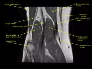
Anatomy • Origin = pubic ramus • Insertion = pesanserinus • Arterial supply • Dominant = terminal branch of MFCA • Minor = branches of SFA and PFA • Venous drainage = venae commitantes • Nerve • motor = anterior branch of obturator nerve • Enters 1-2 cm superior to vascular pedicle • Sensory = medial cutaneous nerve of thigh
Flap variations: • Can harvest with skin paddle (i.e. TUG flap) • Common second choice for breast FTT • +/- innervated
Skin markings • Axis of flap is line from ischium to medial condyle of knee • Or palpate adductor longus: gracillis is 2-3 finger breadths posterior • pedicle marked 10cm below ischium
Flap dissection • Incise over axis of muscle proximally • Optional distal incision to disinsert distal insertion • Dissect down to fascia over gracillis and adductor longus until septum reached • Retract these muscles apart from each other • Pedicle lies here • Proximal origin divided, pedicle dissected