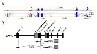
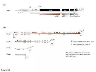
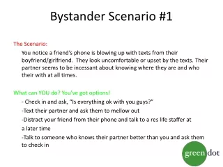
Download
1 / 32
320 likes | 472 Views
Exon-level transcriptome analysis of HIV-1 infected and bystander primary CD4+ T lymphocytes. Michael Imbeault Laboratory of Dr. Michel J. Tremblay Universit é Laval, Québec Canada AIDS 2010 Vienna. Goal.

E N D
Exon-level transcriptome analysis of HIV-1 infected and bystander primary CD4+ T lymphocytes Michael Imbeault Laboratory of Dr. Michel J. Tremblay Université Laval, Québec Canada AIDS 2010 Vienna
Goal • Describe the transcriptomic profile of primary CD4+ T cellsexposed to HIV-1 in vitro • Compare infectedcells and bystandercells
Infected (10%) Problem Mock Control • Quantification of RNA - 10 copies vs 12 copies = 1.2 fold • But in infectedcells, its a 3 fold induction
NL4-3-IRES-HSA • Express all viral genes • Allow for separation of infectedcellsusingmagneticbeads • More sensitive than the parental GFP virus (Levy & al) • Detailspublished in Virology. 2009 Oct 10;393(1):160-7
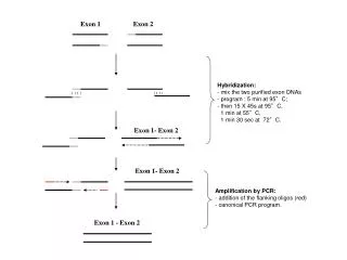
Human Exon 1.0 ST array • Latest offering from Affymetrix • 1.4 millions probesets • 1 million exons • Covers • All knownhumangenes • in silicopredictedgenes • ESTs • Allow for quantification of expression and determination of alternative splicingevents
Separation of infectedcells • Isolate a mean of 500 000 infectedcellsstartingfrom • 50 million cellsatday 1 • 25 million cellsatday 2 and 3 • Extraction of RNA using a dual Trizol – Qiagen custom protocol • Quantification of purity by Taqman qRT-PCR against Tat-spliced
Analysis • Strict statististicalanalysisusingBioconductor’s LIMMA • FDR 1% • 1.7 fold minimum
Automatedlitteratureanalysis • Automatedanalysis of literatureusingGenomatixBibliosphere • Citation of 2 genes in the same sentence in at least 3 different abstracts • Exported to graph management software Gephi • Gephi.org
Main features • AP-1 (FOS and JUN, someotherrelatedgenes) • A group of genesrelated to activated / effector T cells • Many cytokines associated to Th1, Th2, Th17 • Th17 relatedgenes have higher surexpression values, perhapsindicating a highersusceptibility • p53 relatedgenes
FOS JUN AP-1
Cytokine related • Th1 (IFNG, TNF-a, TNF-b, IL1A, IL3) • Th2 (IL4, IL5, IL-10, IL13) • Th17 (IL17A, IL17F, IL21, IL22, IL23R, IRF4)
IL17A IL17F Th17 related genes
IL22 IL23R Th17 related genes
Alternative splicinganalysis • Used the combination of two of the best algorithms • MADS and PECA-SI • P < 0.01 • Splicing index of at least 0.6 • Exon canbedetected in at least 1/3 of arrays • Filter out exons not currentlyassociatedwithgenes
Alternative splicing results • 547 probes in 372 transcripts • 52% of these are in non-codingUTRs • 48% in coding exons
Conclusion • Infectedcells have a transcriptomic profile of highlyactivated / effector / memory T cells • No effectat all in bystandercells • Confirmed a lot of things • Role of p53 in HIV-1 pathogenenis • Manyinteresting candidates • Potentialsusceptibility and restriction factors
Acknowledgements • Michel J. Tremblay • Project Leaders • Corinne Barat, Ph.D. • RéjeanCantin, Ph.D. • Robert Lodge, Ph.D. • Michel Ouellet, Ph.D. • Postdoctoral Fellows • RavendraGarg, Ph.D. • Pranav Kumar, Ph.D. • Guadalupe Andreani, Ph.D. • Masayuki Fujino, Ph.D. • Sandra Côté, Ph.D. • Ph.D. Students • RémiFromentin, M.Sc. • Alexandra Lambert, M.Sc. • Lise-AndréeGobeil, M.Sc. • Jonathan Bertin, M.Sc. • Pascal Jalaguier, M.Sc. • AnissaCheikh, M.Sc. • Alejandro Martin Gomez Lic. • M.Sc. Students • Alexis Danylo, B.Sc. • KatiaGiguère, B.Sc. • Audrey Plante, B.Sc. • Jean-François Bolduc, B.Sc. • VéroniqueVeillette, B.Sc.