Download

1 / 36
500 likes | 816 Views
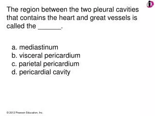
Pericardium & Heart. Dr. Zeenat Zaidi. Heart enclosed within the pericardial sac is located in the middle mediastinum. Mediastinum. The cavity of thorax is divided into: A median partition, the mediastinum Laterally placed pleurae & lungs

E N D
Pericardium & Heart Dr. Zeenat Zaidi
Heart enclosed within the pericardial sac is located in the middle mediastinum
Mediastinum • The cavity of thorax is divided into: • A median partition, the mediastinum • Laterally placed pleurae & lungs • Mediastinum is a thick mobile partition, formed by the structures occupying the central part of the thoracic cavity, between the lungs • It extends: • Superiorly to the thoracic inlet & the root of neck • Inferiorly to the diaphragm • Anteriorly to the sternum • Posteriorly to the twelve thoracic vertebrae
Mediastinum: Divisions • Divided, by an imaginary plane passing from the sternal angleanteriorly to the lower border of the body of the 4th thoracicvertebraposteriorly, into: • Superior mediastinum • Inferiormediastinum • The inferior mediastinum is further divided into: • Anterior • Middle • Posterior
Pericardium • Double layered fibroserous sac that encloses the heart & the roots of the great vessels • Located in the middle mediastinum • Posterior to the body of sternum and 2nd-6th costal cartilages • Anterior to T5-T8 vertebrae • 1-1.5 cm to the right of the sternum • 5-7.5 cm to the left of median plane at the level of 5thintercostal space
Pericardium cont’d • The strong outer layer, the fibrous pericardium is composed of tough fibrous tissue • The inner transparent membrane, the serous pericardium, has two layers: • Parietal • Visceral
Fibrous Pericardium • Tough conical outer fibrous sac, protects heart against sudden overfilling • Superiorly: • Pierced by aorta, pulmonary trunk, and superior vena cava • Becomes fused with the tunica adventitia of these vessels • Below: rests on and is fused with the central tendon of the diaphragm
Fibrous Pericardium cont’d • Anteriorly attached to the posterior surface of sternum by condensations of connective tissue called the sternopericardial ligaments • Posteriorly: • Pierced by pulmonary veins and inferior vena cava • Becomes fused with the tunica adventitia of these vessels
Serous Pericardium • Parietal layer lines the fibrous pericardium and becomes continuous with the visceral layer around the roots of great vessels • Visceral layer reflected onto the heart, forms the external layer of the heart wall (epicardium) • The two layers are continuous with each other at the base of the large vessels • Pericardial cavity lies between the two layers, that contains a thin film of serous fluid which helps in frictionless movement of the heart
Pericardial Sinuses • Develop during folding of embryonic heart • Transverse sinus: • Lies posterior to ascending aorta and pulmonary trunk, anterior to superior vena cava • Communicates with the main part of pericardial cavity at its right and left ends • Oblique sinus: • An inverted U-shaped blind recess lies posterior to the heart extending posterior to the left atrium, can be entered inferiorly • Produced by the reflection of pericardium onto the pulmonary veins and inferior vena cava
Blood Supply • Arterial Supply: • Fibrous pericardium & the parietal layerof the serous pericardium : Mainly supplied by pericardiophrenic and musculophrenic arteries, branches of internal thoracic. Also supplied by pericardial branches of bronchial, esophageal and superior phrenicarteries • Visceral layer of the serous pericardium (epicardium) supplied by the branches of the coronaryarteries • Venous drainage: Veins are tributaries of azygos system. Pericardiophrenic veins also drain into the internal thoracic vein
Nerve Supply • The fibrous pericardium and the parietal layer of the serous pericardium are supplied by the phrenic nerves. • The visceral layer of the serous pericardium is innervated by sympathetic & parasympathetic fibersthrough the sympathetic trunks and the vagus nerves respectively
Clinical Notes • Pericarditis & pericardial effusion • Cardiac temponade • Friction rub • Pericardiocentesis: A wide-bore needle may be inserted through the left 5th & 6th intercostal space near the sternum (area of cardiac notch). Intracardial injections are also given through this area
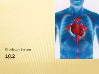
Heart To be handled with care….
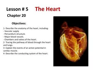
Heart • Hollow muscular organ, acts as a double pump • Conical in shape • Slightly larger than the clenched fist • Lies free within the pericardium • Connected superiorly to the large vessels
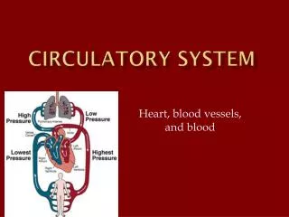
Heart cont’d • Has 4 chambers: two atria superiorly and two ventricles inferiorly, separated from each other by atrioventricular and interventricular grooves • Atria act as the receiving chambers and the ventricles as the pumping chambers • Right side of the heart contains deoxygenated blood & left side contains oxygenated blood
External Features: Surfaces The heart has: • Sternocostal (Anterior) surface • Diaphragmatic (Inferior) surface • Base (Posterior surface) • Apex
Sternocostal Surface • Formed mainly by the right atrium and the right ventricle separated by vertical atrioventricular groove • The anterior interventricular groove separates the right ventricle from the left ventricle
Diaphragmatic Surface • Formed mainly by the right and left ventricles separated by posterior interventricular groove, and a small part of the right atrium, into which the inferior vena cava opens
Base (Posterior Surface) • Formed mainly by the left atrium, into which open the 4 pulmonary veins • Quadrilateral in shape • Lies opposite the apex • Faces posteriorly, superiorly and toward the right shoulder
Apex • Formed by left ventricle • Directed downward, forward and to the left • Located posterior to the left 5thintercostal space, 7-9 cm from the median plane and just medial to the left midclavicular line • Position varies slightly with the person’s position and the phase of respiration • Is the point of maximal pulsation of the heart (the apex beat can be seen as well as palpated)
Borders of the Heart The heart has 4 borders: • Right: Formed by the right atrium • Left: Formed by the left auricle above and the left ventricle below • Inferior: Formed mainly by the right ventricle, and the apex of the left ventricle • Superior border is where great vessels enter or leave the heart. Formed by right and left auricles and superior part of right & left ventricle
Arterial Supply • Supplied by right & left coronary arteries • Coronary arteries arise from the ascending aorta immediately above the aortic valves • Coronary arteries and their branches are distributed over the surface of the heart lying within the subepicardial connective tissue
Right Coronary Artery • Arises from anterior sinus of ascending aorta • Runs between the pulmonary trunk and right auricle • Runs in the atrioventricular groove • At the inferior border of the heart turns posteriorly in the atrioventricular groove • Anastomoses with the left coronary artery in the posterior interventricular groove
Right Coronary Artery: Branches • Right conus artery • Anterior ventricular branches: 2-3 in number, largest is the marginal branch • Posterior ventricular branches, gives a branch to atrioventricular node • Posterior interventricular artery • Atrial branches, & artery of the sinuatrial node which also supplies atria
Left Coronary Artery • Larger than the right • Supplies major part of the heart • Arises from the left posterior aortic sinus • Runs between the pulmonary trunk and left auricle • Runs in the atrioventricular groove • Divides into anterior interventricular & circumflex branches
Left Coronary Artery: Branches • Anterior interventricular artery, gives a small conus artery • Circumflex artery, gives: • Left marginal • Anterior ventricular • Posterior ventricular • Atrial branches
Variations in Coronary Arteries • In 35% of individuals, the sinuatrial artery arises from left coronary artery • In most of the individuals (90%), the posterior interventricular artery is a branch of right coronary artery (Right Dominance). In 10% of the individuals, it arises from circumflex branch of left coronary artery (Left Dominance)
Coronary Artery Anastomoses • Though anastomoses do exist between the terminal branches of the right and left coronary arteries, but these are not large enough to compensate for any sudden blockage of a large branch. • A sudden blockage of the larger branches results in myocardial infarction.
Venous Drainage • Most of the blood drains into the right atrium through the coronary sinus, which: • Lies in the posterior part of the atrioventricularsulcus • Is continuation of the great cardiac vein • Opens into the right atrium to the left of the inferior vena cava Tributaries: • Small cardiac vein • Middle cardiac vein • Posterior vein of the left ventricle • Oblique vein of the left atrium
Venous Drainage cont’d • Anterior cardiac veins drain directly into the right atrium • Vena cordisminimi (Thebesian veins) open directly into heart chambers