Download
1 / 24
240 likes | 361 Views
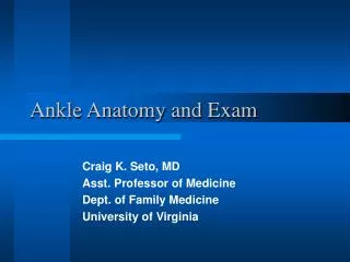
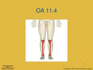
Chapter 19: The Ankle and Lower Leg. Functional Anatomy. Ankle is a stable hinge joint Medial and lateral displacement is prevented by the malleoli Ligament arrangement limits inversion and eversion at the subtalar joint Square shape of talus adds to stability of the ankle

E N D
Functional Anatomy • Ankle is a stable hinge joint • Medial and lateral displacement is prevented by the malleoli • Ligament arrangement limits inversion and eversion at the subtalar joint • Square shape of talus adds to stability of the ankle • Most stable during dorsiflexion, least stable in plantar flexion
Degrees of motion for the ankle range from 10 degrees of dorsiflexion to 50 degrees of plantar flexion • Normal gait requires 10 degrees of dorsiflexion and 20 degrees of plantar flexion with the knee fully extended • Normal ankle function is dependent on action of the rearfoot and subtalar joint
Preventing Injury in the Lower Leg and Ankle • Achilles Tendon Stretching • A tight heel cord may limit dorsiflexion and may predispose athlete to ankle injury • Should routinely stretch before and after practice • Stretching should be performed with knee extended and flexed 15-30 degrees • Strength Training • Static and dynamic joint stability is critical in preventing injury • While maintaining normal ROM, muscles and tendons surrounding joint must be kept strong
Footwear • Can be an important factor in reducing injury • Shoes should not be used in activities they were not made for • Preventive Taping and Orthoses • Tape can provide some prophylactic protection • However, improperly applied tape can disrupt normal biomechanical function and cause injury • Lace-up braces have even been found to be superior to taping relative to prevention
Assessing the Lower Leg and Ankle • History • Past history • Mechanism of injury • When does it hurt? • Type of, quality of, duration of pain? • Sounds or feelings? • How long were you disabled? • Swelling? • Previous treatments?
Percussion and compression tests • Used when fracture is suspected • Percussion test is a blow to the tibia, fibula or heel to create vibratory force that resonates w/in fracture causing pain • Compression test involves compression of tibia and fibula either above or below site of concern • Thompson test • Squeeze calf muscle, while foot is extended off table to test the integrity of the Achilles tendon • Positive tests results in no movement in the foot • Homan’s test • Test for deep vein thrombophlebitis • With knee extended and foot off table, ankle is moved into dorsiflexion • Pain in calf is a positive sign and should be referred
Compression Test Percussion Test Homan’s Test Thompson Test
Ankle Stability Tests • Anterior drawer test • Used to determine damage to anterior talofibular ligament primarily and other lateral ligament secondarily • A positive test occurs when foot slides forward and/or makes a clunking sound as it reaches the end point • Talar tilt test • Performed to determine extent of inversion or eversion injuries • With foot at 90 degrees calcaneus is inverted and excessive motion indicates injury to calcaneofibular ligament and possibly the anterior and posterior talofibular ligaments • If the calcaneus is everted, the deltoid ligament is tested
Anterior Drawer Test Talar Tilt Test
Functional Tests • While weight bearing the following should be performed • Walk on toes (plantar flexion) • Walk on heels (dorsiflexion) • Walk on lateral borders of feet (inversion) • Walk on medial borders of feet (eversion) • Hops on injured ankle • Passive, active and resistive movements should be manually applied to determine joint integrity and muscle function • If any of these are painful they should be avoided
Specific Injuries • Ankle Injuries: Sprains • Single most common injury in athletics caused by sudden inversion or eversion moments • Inversion Sprains • Most common and result in injury to the lateral ligaments • Anterior talofibular ligament is injured with inversion, plantar flexion and internal rotation • Occasionally the force is great enough for an avulsion fracture to occur w/ the lateral malleolus
Syndesmotic Sprain • Etiology • Injury to the distal tibiofemoral joint (anterior/posterior tibiofibular ligament) • Torn w/ increased external rotation or dorsiflexion • Injured in conjunction w/ medial and lateral ligaments • Signs and Symptoms • Severe pain, loss of function; passive external rotation and dorsiflexion cause pain • Pain is usually anterolaterally located • Management • Difficult to treat and may requires months of treatment • Same course of treatment as other sprains, however, immobilization and total rehab may be longer
Achilles Tendon Rupture • Etiology • Occurs w/ sudden stop and go; forceful plantar flexion w/ knee moving into full extension • Commonly seen in athletes > 30 years old • Generally has history of chronic inflammation • Signs and Symptoms • Sudden snap (kick in the leg) w/ immediate pain which rapidly subsides • Point tenderness, swelling, discoloration; decreased ROM • Obvious indentation and positive Thompson test • Occurs 2-6 cm proximal the calcaneal insertion
Achilles Tendon Rupture (continued) • Management • Usual management involves surgical repair for serious injuries (return of 75-80% of function) • Non-operative treatment consists of RICE, NSAID’s, analgesics, and a non-weight bearing cast for 6 weeks, followed up by a walking cast for 2 weeks (75-90% return to normal function) • Rehabilitation lasts about 6 months and consists of ROM, PRE and wearing a 2cm heel lift in both shoes
Medial Tibial Stress Syndrome (Shin Splints) • Etiology • Pain in anterior portion of shin • Catch all for stress fractures, muscle strains, chronic anterior compartment syndrome • Accounts for 10-15% of all running injuries, 60% of leg pain in athletes • Caused by repetitive microtrauma • Weak muscles, improper footwear, training errors, varus foot, tight heel cord, hypermobile or pronated feet and even forefoot supination can contribute to MTSS • May also involve, stress fractures or exertional compartment syndrome
Shin Splints (continued) • Signs and Symptoms • Four grades of pain • Pain after activity • Pain before and after activity and not affecting performance • Pain before, during and after activity, affecting performance • Pain so severe, performance is impossible • Management • Physician referral for X-rays and bone scan • Activity modification • Correction of abnormal biomechanics • Ice massage to reduce pain and inflammation • Flexibility program for gastroc-soleus complex • Arch taping and or orthotics
Compartment Syndrome • Etiology • Rare acute traumatic syndrome due to direct blow or excessive exercise • Signs and Symptoms • Excessive swelling compresses muscles, blood supply and nerves • Increase in fluid accumulation could lead to permanent disability • Chronic cases appear as gradual build-up that dissipates following activity; generally bilateral and becomes predictable; can remain elevated producing ischemia and pain or ache w/ rare neurological involvement; increased pressure involvement • Weakness with foot and toe extension and occasionally numbness in dorsal region of foot
Compartment Syndrome (continued) • Management • If severe acute or chronic case, may present as medical emergency that requires surgery to reduce pressure or release fascia • RICE, NSAID’s and analgesics as needed • Surgical release is generally used in recurrent conditions • Return to activity after surgery - light activity- 10 days later
Functional Progressions • Severe injuries require more detailed plan • Introduction of weight bearing activities (partial vs. full) is critical to progress • Progression must occur based on pain and level of function • Running can begin when ambulation is pain free (transition from pool even surface changes of speed and direction)
Return to Activity • Must have complete range of motion and at least 80-90% of pre-injury strength before return to sport • If full practice is tolerated w/out insult, athlete can return to competition • Return to activity must involve gradual progression of functional activities, slowly increasing stress on injured structure • Specific sports dictate specific drills