Download

1 / 38
380 likes | 443 Views
Follow the Signs:. Advanced thinking in a patient encounter Amanda Waggoner, COMT. Objectives. -Become more efficient technicians -Identify information and testing needed to BEST help the patient and doctor -Streamline exam time. STOP.

E N D
Follow the Signs: Advanced thinking in a patient encounter Amanda Waggoner, COMT
Objectives • -Become more efficient technicians • -Identify information and testing needed to BEST help the patient and doctor • -Streamline exam time
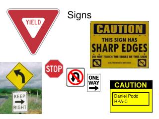
STOP • Remember to stop before each patient and take a breath to prepare for a new encounter. • Don’t assume because a patient is scheduled for a specific type of exam that is why they are here. • Take a moment to review their past history to see if any testing was requested for the next exam and to familiarize yourself with their history. • Be Proactive-If the patient has a specific problem and regular testing is indicated and overdue take action (test same day, alert Physician, schedule for another date).
LISTEN • Listen to what the patient is complaining of and clearly state this in the record. • Try to stay out of a “rut” of common complaints (Blurred vision, red eye, etc.) Use the patients words when documenting complaint. • Realize some patients will not want to complain, while others will tell you everything. Try to guide the patient to focus on what is their main reason for being there. • Repeat their complaint as you are entering it to make sure you are “hearing” them correctly.
Chief Complaint • The chief complaint is your guide. • Remember to ask questions about complaints, a general “floaters” could be a number of problems. As technicians WE can make the Physicians job easier when learning to recognize signs that lead us to proactively streamline an exam and save time.
Floaters Posterior Vitreous Syneresis Ophthalmic migraine Light or dark colored “spots” in vision. Move with gaze and if patient tries to focus on them. More noticeable in Bright light. How many? Changing Shape? Any flashes of light? Light pattern, described as (lightning shaped, heat wave, circular pattern). Usually only lasting 10 min-30min, may or may not be followed by a headache. May start peripherally and move central. Symptoms can be OU.
Loss of Vision Blurred Vision “Gone” vision Blurriness, Glare. Gradual or sudden? With glasses or without? Near vision or distance? Associated pain or redness with onset? Blanked out or darkened out area’s. Sudden or gradual? Distortion in vision? Health history can be a big clue. Recent history of “bumping” into things on a side, or trouble judging space when parking?
Red Eye Subconjunctival hemorrhage Iritis Engorged red veins. Patients may also complain of decreased vision, light sensitivity, and aching. • Blood red, completely covering an area. May have started small but grown in size. Other people may have noticed first. No change in vision. No pain or only slightirritation.
Pain Irritation/ Foreign body Achiness/Soreness Duration? Location? Change in vision? Light sensitivity? Associated headache or nausea? • Duration? What helps relieve symptoms? Injury or Chemical exposure? CL wearer? Tearing or discharge?
Double Vision • Monocular-not a muscle issue. • Any distortion? Think retina. • Double not “real” images only with lights? Think media opacity. • Vision described as elongated or skewed? Think refractive error. • Binocular-goes away with one eye closed. • All the time? If not, when do symptoms start? Near, Distance or both? • Sudden onset or worsening over time? • Any associated lid droop or pupil changes noted?
LOOK • Observe the patient while taking histories. -Redness and swelling -crusting or discharge on lids -proptosis of one or both eyes -Eyelid closing or guarding of an eye -abnormal head positioning -facial or eyelid droop Make note on chart
Exam-Visual Acuity Testing strategies-Snellen/Allen/Tumbling E’s/numbers use what is easiest for the patient to understand -Pinhole-Use when BCVA is lower than expected Special Cases Language barriers/ Nystagmus/ “surprise loss of vision”
EOM and Cover Testing • Test with glasses so patient can fixate at distance/near easily. Position correctly so patient is using the right part of glasses. • Check Corneal reflexes-I use a pen light while doing EOM’s so I can “double check” • Never rely on past exam findings-Did the last Tech miss it? Has the patient developed a palsy since last exam? ONLY RECORD IN A CHART WHAT YOU HAVE CHECKED YOURSELF! • ALWAYS have the doctor check patient before dilation with new onset or change in diplopia that is constant or frequent.
EOM and Cover Testing • Our job as technicians is to recognize anything “abnormal. Do not worry if you can’t specify the exact prism diopter needed. The physicians will have more trust in you if you notice a problem and have them check, than if you ignore it because you are nervous about getting the “right” answer. • If everything is ortho on exam but you pick up Prism in the glasses, recheck without glasses. Patients do not always know to tell us because with glasses VA is fine. If no diplopia with current, Great! We still need notation of why it is needed.
EOM and Cover Testing • EOM-What are we checking? • Use a Square or Star shape. • Make sure you are extending far enough in each direction for the patient to reach endgaze. • If the patient has difficulty following your fixation device you can verbally ask them to look in each direction. • Do not be afraid to lift a lid if needed, or check for corneal reflex in different positions.
EOM and Cover Testing • We are not Magicians…avoid the magic wand! • Determine Tropia vs. Phoria Cover/Uncover to determine Tropia Alternate Cover test to determine Phoria -Check both ways!
Confrontation Visual Fields • We are not performing a full visual field! This is a screening evaluation to pick up gross loss. • Why is it important? Retinal vascular problems, Optic Nerve dysfunction, Stroke, Brain Tumor…Big stuff! • We are testing against our own assumed normal visual field, cover your opposite eye so the target is seen simultaneously by the patient and you. • If VA is poor use a light, a bright colored medicine cap or hand motion can be used for a target.
Visual Pathway When doing Confrontation visual fields, know your Visual Pathway. This is a “map” to where any dysfunction is stemming from. Prechiasmal-Monocular defects Chiasmal- Heteronymous defects-the chiasm is the only place nasal fibers from both eyes can be affected. Postchiasmal-Homonymous defects-The more congruent (same) the defects are, the more posterior in the brain the lesion is located.
Pupils • Always check before dilation! If unsure get a double check. • Do not assume the last technician was correct. If the patient has an Optic Nerve condition or poor vision suspect possible APD and always check. • If no new complaints and 20/20 VA, APD is unlikely. • Check for subjective APD-ask the patient which eye is the light brighter in, this can be found even in early cases of Optic Neuropathy where an APD may not yet be completely visible.
Pupils • Anisocoria? <1 mm difference in pupils is considered anatomically normal. • Any Trauma to either eye (past or new), Surgery on one or both eyes?, History of inflammation (iritis)? • Is one eye dilated? Has the patient been exposed to any chemicals, are they using a redness reliever frequently, have they used a motion sickness patch, is it associated with a migraine? • Is one eye miotic? Any associated symptoms of lid droop or eye turn? Does the pupil react to convergence (assess while patient focuses on small print).
Refractometry • Where do I start? Lensometry/ Last MR on record/Autorefraction/Retinoscopy • Do not forget to check for cylinder, if no previous Rx available or autorefractor start with option of .50 at both main and oblique. • Balance changes in cyl and sphere. • Use your tools: Fogging/ Red-Green • Does complaint “match” RX? Latent Hyperopes, Cataract changes, Diabetic changes?
Refractometry • Handling the patient -”I hate this test, I can never decide” -”you are not giving me enough time” -” I like choice 2, from 5 min ago best” -”shouldn’t the Doctor be doing this?”
Refractometry • ADD power, Does it make sense??? Preferred position of material. Special Circumstances • Low vision (eccentric fixation, rolling spheres, aphakia, high ADD). • Trial frame MR (Tremors, head positioning or physical disability). • Illiterate or Developmentally disabled patients. • Vertex and do quick OR with current glasses • Be Flexible and creative.
Slit Lamp • We are not “just” checking pressure. • Always do a quick sweep of each eye to determine any abnormalities. • Check with white light-lids and lash margins/ Cornea/ Anterior Chamber depth/ iris • Check under blue filter with fluorescein- Tear film break up time and any staining. • Perform Applanation tonometry • RECHECK cornea! If you catch a lash or abrade the cornea you want to know before the doctor finds it!!!!
Tonometry • Always check your equipment. Is the tonometer prism clean? Are the marks aligned at 180? Make sure when swinging into position, everything “clicks” into setting. • Remind the patient to keep their mouth shut (this is really the only time we can get away with this), breathe normally, teeth together, and BOTH eyes open. • If you need to hold a lid put minimal pressure against the globe, try to use the brow bone instead. • CLEAN YOUR TONOTIP after use, and swing tonometer back to the “resting” position.
Tonometry • If a value is outside of normal and you are unsure of reading, recheck. • This is where Histories can be a help. Is the patient on Glaucoma Therapy and are they compliant? Are they using any steroids (ocular or systemic), Do they have Thyroid problems (check in straight gaze and upgaze), are they postsurgical? • If reading is high and has always been high normal-are their angles open? Has Pachymetry ever been done? • Do not proceed with IOP if possible Shingles/HSK. If there are any corneal complaints check eye before proceeding with drops and IOP reading. It is OK to defer and ask before “touching” an eye.
Dilation • Check for drop protocol when working in unfamiliar clinics. • ALWAYS check your labels before instilling drops. • If Latent Hyperopia is suspected or possible use a Cycloplegic drop. • Explain to the patient vision will be affected and that they will need sunglasses. • If the patient complains of previous “allergy” get specifics on reaction and ask the physician how to proceed.
When do I not use drops? No drops before Physician OK DO NOT DILATE If prism needs to be assessed. If the patient presents with a possible Neurological problem and there is a possible APD. If there are any iris abnormalities that need to be measured or seen by physician. If narrow angle suspected. • Suspected HSK/Shingles • Foreign body or trauma injuries where Penetration of anterior chamber is possible. • Post surgical for Ruptured globe, large corneal or scleral incision repair, refractive surgery by protocol.
Testing • Brightness Acuity Testing -glare complaints -History of Cataract or possible PC haze. Check if visual acuity is better than 20/50 on any Cataract eval. Insurance requires for qualification to proceed with surgery. • Pachymetry -High normal-high IOP -Glaucoma evaluations -Peripheral cornea if surgeon planning a manual LRI -Corneal Dystrophies Be proactive, if not in chart go ahead and check with Glaucoma/ Ocular Hypertension.
Testing • Amsler -distortion complaints -Flashes/Floaters -monocular diplopia -screening with cataract evaluations -High Risk Medication patients • Color Testing -Kids -Multiple Sclerosis patients -High Risk Medication patients -Optic Nerve/ Neuro -Traffic Lights for DMV
Testing • Hertel / exophthalmometer -Thyroid Eye Disease -Traumatic injuries that may involve orbit. -Scleral show or Proptotic appearing eyes. • Corneal Topography -Pre or Post LASIK/RK patients -Post PKP patients -irregular astigmatism with decreased best corrected vision -History of Kerataconus
Testing • OCT-Macula -Post Surgical with decreased vision -High risk Medication patients -Change in Amsler grid -loss of central vision or complaints of micropsia/macropsia • OCT-Optic Nerve -Glaucoma -Ocular Hypertension -Optic Neuritis/ Ischemia -Change in color discrepancy between eyes
Conclusion • Think while doing the exam- What could this be? What can I do to rule in or out any possibilities? Is there a finding I am uncomfortable making a firm decision on? • Do not be afraid to ask for help. • Be proactive. Is there information you can give the patient to facilitate their understanding of a condition? • Review the charts after doctor has seen patient to see what the outcome was, use this as a learning tool. • Have PASSION for your job, always keep learning!