Download

1 / 19
250 likes | 1.19k Views
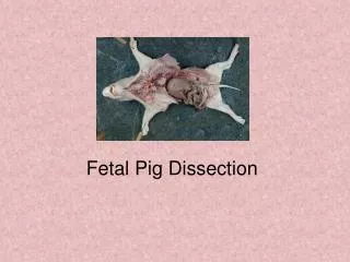
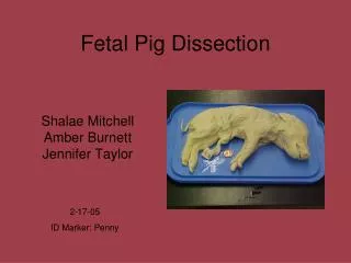
Fetal Pig Dissection. Shalae Mitchell Amber Burnett Jennifer Taylor. 2-17-05 ID Marker: Penny. The fetal pig our group is working on was not killed for this. It was found in a sow marked for slaughter, and was never born. It was then given to us for use in science.

E N D
Fetal Pig Dissection Shalae Mitchell Amber Burnett Jennifer Taylor 2-17-05 ID Marker: Penny
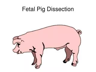
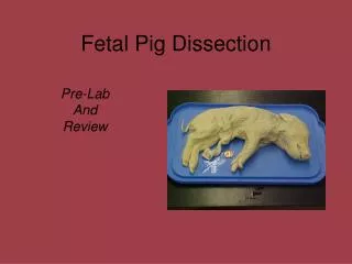
The fetal pig our group is working on was not killed for this. It was found in a sow marked for slaughter, and was never born. It was then given to us for use in science. • Sus scrofa is the technical name of the domestic pig. They eat both plants and animals. Their body temperature is slightly higher than humans. They live 15 to 20 years. Pigs are often used in experiments on human drugs. There is a new “mini-pig” that weighs 100-150 lbs. especially for that use. • During gestation, there is an exchange of substances between the blood of te mother and of the embryo across the placenta through the umbilical cord.
Our pig is 34 cm long, and was 112-115 days old at birth, which is full term.
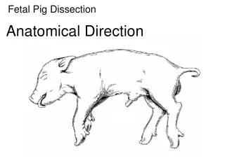
Ventral (inferior) Posterior (caudal) Anterior (cranial) Anatomical Directions Lateral Median Sagittal Transverse Frontal (coronal) Distal Dorsal (superior) Proximal
Lumbar region Mammilary papillae tail ankle Umbilical cord
Our fetal pig is male. How many pairs of mammary papillae do you count? 7 ½ pairs Do all specimens in class have the same amount? No
Pig Toes Only the 2nd, 3rd, and 4th digits touch the ground. The 1st is nonexistent and the 5th never touches it. 2nd/3rd Digits 4th Digit
Pig Dissection Notes • Use scalpel sparingly • Rely primarily on your dissecting needles. • Leave organs intact unless directed to do otherwise. • When using scissors, walk w/the blunt end out. • Move organs aside w/your fingers or a blunt probe. • Apply lanolin or Vaseline at the outset or wear thin rubber gloves. • Line your dissection pan w/paper towels in order to absorb excess fluids, as a storage for structures removed, and to ease cleaning up at the end. • Wrap the fetal pig in wet paper towels before returning it to the bag. • Twist the top of the bag and close tightly w/a rubber band. • Apply the following solution w/a one inch paint brush at the close of each session to preserve the softness and texture of the pig: 30 grams of Carbolic Acid crystals, 250 ml. of Glycerin, and 1000ml of Water.
Salivary Glands Masseter Muscle Parotid Gland Facial Nerve, Dorsal Branch Facial Nerve, Ventral Branch Submaxillary Gland External Maxillary Vein Stensen’s (Parotid) Duct.
Teeth 3/3 Incisors 1/1 Canines 4/4 Premolars 0/0 Molars
Nostril Oral Cavity Hard Palate Soft Palate Epiglottis Opening to Naso-Parynx Corner of Mouth (cut surface) Tongue Papillae of Tongue
Canine Tooth Hard Palate Corner of mouth Soft Palate Opening to Naso-Pharynx Opening to Trachea Mandible Epiglottis
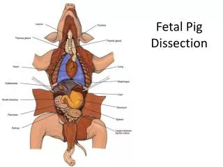
Stomach Liver L. I. S. I. Umbilical Artery
L.I. Liver Umbilical Artery S.I. Urinary Bladder Kidney, Spleen, Ceacum, and Stomach not shown
Stomach Pancreas L.I. Liver S. I.
Human Digestive System
Page 56, #1 – 9 • Divisions of pharynx: nasopharynx, oropharynx, and the laryngopharynx/hypopharynx. • 7 passages that penetrate the pharynx: mouth and nasal passages, Eustachian tubes, larynx, esophagus, glottis, and pharyngeal area, • 4 types of papillae found on the surface of the tongue: sweet, sour, bitter, salty. Differences: They react to different molecules of food. • Bones of the hard palate: palatine rugae • Other than the palatine tonsils, the other tonsils are: lingual and pharyngeal. • Secretions from the salivary glands begin the digestion of : food • The esophagus is located: c) dorsal to the trachea • A major blood vessel that is formed just posterior to the submaxillary gland as a result of the union of smaller vessels is external maxillary gland is the submental branch of the Facial artery (off of the External Carotid artery) • The cap of cartilage which prevents food from entering the trachea while swallowing is the epiglottis.
Page 66 #1-9 1. Lobes of the Liver: left, right, lateral, medial 2. Structures of the greater and lesser omentum:greater is from above stomach and below transverse colon and the lesser is covers and supports various organs and blood vessels. 3. Jejunum:8 ft. long Ileum: 12 ft. long 4. Location and function on ileocecal valve: separates the last part of the small intestine and the first part of the large intestine keeps food in the S. I. until digestion is complete. 5. Parts of the alimentary canal: mucosa, submucosa, muscularis, and the adventitia. 6. *Location of the adrenal gland: Pig - above kineys Human - on top of kidneys. 7. Digestion of fats begin in the small intestine. 8. 3 major tubular structures in the diaphragm: the aorta, the esophagus and the posterior vena cava 9. The trachea is not in the abdominal cavity.
Computer: Jennifer Taylor Photographer: Shalae Mitchell Dissector: Amber Burnett