Download
1 / 23

E N D
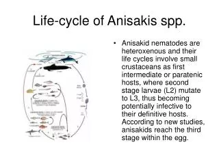
Anisakid nematodes are heteroxenous and their life cycles involve small crustaceans as first intermediate or paratenic hosts, where second stage larvae (L2) mutate to L3, thus becoming potentially infective to their definitive hosts. According to new studies, anisakids reach the third stage within the egg. Life-cycle of Anisakis spp.
Eukaryota Metazoa Nematoda SecernenteaAscarididaAscaridoideaAnisakidae The most frequently recovered anisakid species in fish belong to the following genera: Anisakis, Pseudoterranova, Contracaecum, Phocascaris Taxonomy
Anisakid larvae can be identified at genus level by light microscopy, mainly on the basis of the morphology of digestive tract and of excretory system.
The taxonomy of anisakids is mainly based on the morphology of male adult specimens. The most significant structural characters for species identification are the distribution and pattern of caudal papillae, the spiculae and the morphology of cephalic end (Fagerholm, 1991).
However, anisakid nematodes tend to be very conserved in gross morphology and molecular techniques have shown that many presumed monospecific species consists of several cryptic species (Nascetti et al., 1993; Paggi et al., 1991; Orecchia et al., 1994; Mattiucci et al., 1997). • Molecular markers in the ribosomal DNA spacers have been recently established for the correct identification of Anisakis cryptic and morphospecies, irrespective of their sex or life history stage (D’Amelio et al., 2000)
1 A.pegreffii2 A.simplex s.s.3 A.simplex C4 A.physeteris5 A.schupakovi6 A.ziphidarum7 A.typicaL 100 bp ladder
Epizootiology • Anisakid larvae have been detected worldwide in a large variety of fish species. Among teleosts, they have been found in Gadiformes, Perciformes, Clupeiformes, Pleuronectiformes, Scorpaeniformes, Zeiformes, Bericiformes, Lophiiformes, Anguilliformes and Atheriniformes.
Third stage larvae of anisakids are commonly found in the flesh and the body cavity of a large number of fishes as well as in cephalopods that serve as paratenic hosts.
Anisakis sp. larva in the flesh of blue whiting (Micromesistius poutassou) after light candling (from www.usc.es/banim/doc/tppanisa.htm)
Prevalence of infection is usually rather low in crustaceans (about 0.2%), while it can reach 100% in several fish species. This suggests that paratenic hosts play a role of worm burden accumulation in anisakids. • Intensity of infection in fish can be rather high, reaching peaks of more than 250 larvae per fish, for instance in Lepidopus caudatus in the Mediterranean Sea.
Anisakid larvae have been also detected in elasmobranchs and in a variety of cephalopods. Among these, they have been found in Octopodidae, Sepiidae, Loliginidae and Ommastrephidae.Adult forms of Anisakis spp. are usually found in cetaceans (whales and dolphins). Pseudoterranova decipiens matures mainly in pinnipeds (phocids and otariids). Some members of the genus Contracaecum reach maturity in pinnipeds, others in fish-eating birds (e.g cormorants, pelicans and herons). Phocascaris species become adults in pinnipeds of the northern hemisphere.
Histopathological studies in fish revealed damage mostly at the level of stomach wall, liver, gonads and muscles. These include mechanical damage, necrosis, tissue compression and castration. Clinical signs in fish are mainly cellular infiltration, and hemorrhage. Histopathology
Histopathological studies in cephalopods revealed damage mostly at the level of stomach wall, mantle muscle, nidamentary glands, testicle and ovary. These include mechanical damage, necrosis, tissue compression and castration. Cellular infiltration is oftenly found associated with encapsulated larvae.
In the stomach of cetaceans, anisakid adults are often found in clusters of individuals embedded in the mucosa and submucosa. Ulcers of 5x3 cm associated with anisakids are found in the fundic portion of the stomach
Human anisakidosis • Anisakids are of medical and economic significance. Larval forms of anisakid nematodes of the genera Anisakis Dujardin, 1845 and Pseudoterranova Mozgovoi, 1951 are in fact the principal aetiological agents of human anisakidosis. • This fish-borne anthropozoonosis occurs when the larvae are taken alive after the consumption of raw, undercooked, or improperly processed fish or cephalopods that serve as paratenic hosts in the life cycle of these nematodes.
Human anisakidosis is becoming of major health and economic importance and it is particular relevant in countries such as Japan, where the number of human cases reach significant peaks owing to the widespread custom of eating preparations based on raw fish, such as sushi and sashimi. Human cases are increasingly reported from United States and many European countries (UK, France, Italy and Spain). • Clinically, several different types of human anisakidosis have been defined based on the location (gastric, intestinal or extra-gastrointestinal) (Ohtaki and Ohtaki, 1989; Ishikura, 1990; Yoshimura, 1990) and on histopathological classification (Kojima et al., 1966).
http://www.stanford.edu/class/humbio103/parasitepages/ParaSites/anisakiasis/Diag.html)
Recently, larval forms of A. simplex have been identified as responsible for IgE-mediated allergic reactions, with symptoms ranging from urticaria, to asthma up to anaphylactic shock (Audicana et al., 2002).