Download
1 / 63
630 likes | 804 Views
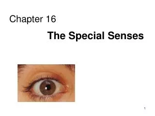
THE SPECIAL SENSES (EYE & EAR). Overview of the Eye. Eye acts much like a camera Lens of eye adjusts to bring object into clear focus Pupil of eye constricts to allow less light to enter in bright setting or dilates to allow more light to enter in darker setting

E N D
Overview of the Eye • Eye acts much like a camera • Lens of eye adjusts to bring object into clear focus • Pupil of eye constricts to allow less light to enter in bright setting or dilates to allow more light to enter in darker setting • Through bending of light rays, image reaches retina • Sensitive nerve cell layer of the eye • Image is transmitted to brain for interpretation
Structures of the Eye(Front View) • Sclera • White portion of the eye • Tough connective tissue • Maintains the shape of the eyeball • Serves as protective covering for the eye • Iris • Colored portion of the eye • Pupil • Opening in the center of the eye • Controls the amount of light entering the eye
Structures of the Eye(Front View) (continued) • Conjunctiva • Thin mucous membrane layer that lines anterior part of eye and inner part of eyelids • Colorless, but appears white because it covers sclera • Lacrimal gland • Located at upper outer edge of each eye • Produces tears • Lacrimal duct • Located at inner edge of eye • Tears drain from eye through this duct
Structures of the Eye(Front View) (continued) Front View of the Eye
Structures of the Eye(Front View) (continued) • Eyelids • Continuous with the skin and cover the eyeball • Keep surface of eyeball lubricated and protected from dust and debris through blinking motion • Eyelashes • Located along the edges of the eyelids • Help protect eyeball by preventing foreign materials and/or insects from coming in contact with surface of eyeball
Structures of the Eye(Cross Section) • Sclera • “White of the eye” • Thinnest over the anterior surface of the eye • Thickest at the back of the eye, near opening for optic nerve • Cornea • Continuous with anterior portion of sclera • Transparent, nonvascular layer covering the colored part of the eye
Structures of the Eye(Cross Section) (continued) • Conjunctiva • Mucous membrane that lines inner surfaces of the eyelids and outer surfaces of the eye • Choroid • Vascular middle layer of the eye • Just beneath the sclera • Contains extensive capillaries that provide blood supply and nutrients to the eye • Contains the iris, ciliary body, and suspensory ligaments
Structures of the Eye(Cross Section) (continued) • Iris • Colored portion of the eye • Can be seen through the transparent corneal layer • Pupil • Located in center of iris • Controls amount of light entering eye • Lens • Colorless biconvex structure that aids in focusing the images clearly on the retina
Structures of the Eye(Cross Section) (continued) • Ciliary Body • Located on each side of the lens • Contains muscles that are responsible for adjusting the lens to view near objects • Suspensory Ligaments • Radiate from the ciliary body and attach to the lens • Hold the lens in place • Assist in adjusting the shape of the lens for proper focusing of the eye
Structures of the Eye(Cross Section) (continued) • Retina • Sensitive nerve cell layer • Changes the energy of the light rays into nerve impulses • Transmits nerve impulses via optic nerve to brain for interpretation of the image seen by the eye • Nerve cells of retina • Rods are responsible for vision in dim light and for peripheral vision • Cones responsible for visualizing colors, central vision, and vision in bright light
Structures of the Eye(Cross Section) (continued) • Retina (continued) • Macula Lutea • Oval, yellowish spot near the center of the retina • Fovea Centralis • Small depression located within the macula lutea • Sharpest image is obtained when image focuses directly on fovea centralis = central vision
Structures of the Eye(Cross Section) (continued) • Optic Nerve • Receives impulses from retina and transmits them to the brain • Images are then interpreted as vision • Optic Disc • Contains no rods or cones • Known as the “blind spot” of the eye • Center of optic disc serves as point of entry for artery that supplies retina
Structures of the Eye(Cross Section) (continued) Lateral Cross Section of the Eye
Structures of the Eye(Cross Section) (continued) • Anterior Cavity of the Eye • Anterior Chamber • Located in front of the lens • Filled with clear, watery fluid called aqueous humor • Posterior Chamber • Located behind the lens • Also filled with aqueous humor – flows back and forth between both chambers
Structures of the Eye(Cross Section) (continued) • Posterior Cavity of the Eye • Posterior to the lens • Filled with vitreous humor • Clear, jellylike substance that gives shape to the eyeball • Not constantly reproduced • Blindness can result if vitreous humor escapes from eye
Process of Vision • Process Begins as Light Rays Enter Eye • Transmitted through cornea, aqueous humor, pupil, lens, and vitreous humor to retina • Sensitive nerve cells of retina transmit image through optic nerve to brain • Brain interprets image as vision
Process of Vision (continued) • Refraction • Process of bending of light rays as they pass through the various structures of the eye to produce a clear image on the retina • Errors of Refraction • Occur when eyeball is abnormally shaped • Occur when lens has lost ability to accommodate to near vision • Vision will be blurred • Can be adjusted with corrective lenses
Astigmatism • Pronounced • (ah-STIG-mah-tizm) • Defined • A refractive error causing the light rays entering the eye to be focused irregularly on the retina due to an abnormally shaped cornea • Correction: contact lenses or eyeglasses, which neutralize the defect
Blepharitis • Pronounced • (blef-ah-RYE-tis) • Defined • Inflammation of the eyelid margins stemming from seborrheic, allergic, or bacterial origin
Blindness • Pronounced • (BLINDNESS) • Defined • The absence of vision or the need for assistive devices and/or assistance from others to accomplish daily activities due to the inability to see
Monochromatism(Color Blindness) • Pronounced • (mon-oh-KROH-mah-tizm) • Defined • An inability to sharply perceive visual colors • Daltonism – unable to distinguish greens from reds • Achromatic Vision – cannot distinguish any color; perceives only white, gray, and black
Cataract • Pronounced • (KAT-ah-rakt) • Defined • Lens in the eye becomes progressively cloudy losing its normal transparency • Thus altering the perception of images due to the interference of light transmission to the retina
Conjunctivitis, Acute • Pronounced • (kon-junk-tih-VYE-tis acute) • Defined • Inflammation of the mucous membrane lining the eyelids and covering the front part of the eyeball • Also called “Pinkeye”
Glaucoma • Pronounced • (glau-KOH-mah) • Defined • Ocular disorders identified as a group due to the increase in intraocular pressure
Glaucoma (continued) • Chronic Open-Angle Glaucoma • Primary disorder • Breakdown in drainage system of the circulation of aqueous humor • Gradual elevation of internal pressure leads to decreased blood supply to optic nerve and retina • Peripheral vision is gradually lost when untreated
Glaucoma (continued) • Acute Closed-Angle Glaucoma • Mouth of drainage system is narrow and closes completely – allowing no flow of aqueous humor • Rapid occurrence characterized by severe pain, blurred vision, photophobia, redness, and seeing “halos” around light • If untreated, person can lose his or her sight within several days
Hyperopia • Pronounced • (high-per-OH-pee-ah) • Defined • A refractive error in which the lens of the eye cannot focus on an image accurately, resulting in impaired close vision that is blurred due to the light rays being focused behind the retina because the eyeball is shorter than normal • Also called farsightedness
Hyperopia (continued) Hyperopia (Farsightedness)
Myopia • Pronounced • (my-OH-pee-ah) • Defined • A refractive error in which the lens of the eye cannot focus on an image accurately, resulting in impaired distant vision that is blurred due to the light rays being focused in front of the retina because they eyeball is longer than normal • Also known as nearsightedness
Myopia (continued) Myopia (nearsightedness)
Presbyopia • Pronounced • (prez-bee-OH-pee-ah) • Defined • A refractive error occurring after the age of 40, when the lens of the eye(s) cannot focus on an image accurately due to its decreasing loss of elasticity resulting in a firmer and more opaque lens • Also called farsightedness due to better clarity of distant objects
Strabismus • Pronounced • (strah-BIZ-mus) • Defined • Failure of the eyes to gaze in the same direction due to weakness in the muscles controlling the position of the eye • Most common type is nonparalytic strasbismus, an inherited defect in which the eye position of the two eyes has no relationship
Strabismus (continued) • Convergent Strabismus • Also known as “Crosseye” • Also known as “Esotropia” • Affected eye turns inward • Usually develops in infancy or early childhood
Strabismus (continued) • Divergent Strabismus • Also known as “Walleye” • Also known as “Exotropia” • Affected eye turns outward
Strabismus (continued) Strabismus:(A) Convergent;(B) Divergent
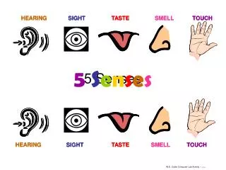
Overview of the Ear • Two important functions of the ear • It enables us to hear • It functions as the sensory organ of balance or equilibrium • Location of one ear on each side of head produces binaural hearing • Hearing from both sides
Structures of the Ear • External Ear • Visible portion of the ear not contained within the head • Auricle or pinna • Cartilaginous flap that has a fleshy, lower portion called the ear lobe • External auditory canal • Tube leading from the auricle to the middle ear • Lined with tiny hairs called cilia which aid in transmitting sound waves inward • Tympanic membrane (eardrum) • Separates external ear from middle ear
Structures of the Ear (continued) • Middle Ear • Contains three tiny bones known as the auditory ossicles • Malleus • Resembles the shape of a hammer • Connected to tympanic membrane and transmits sound vibrations to second auditory ossicle • Incus • Resembles the shape of an anvil • Transmits sound vibrations from malleus to third auditory ossicle
Structures of the Ear (continued) • Middle Ear (continued) • Stapes • Shaped like a tiny stirrup • Transmits sound vibrations from incus to the inner ear • Eustachian tube • Connects the middle ear to the pharynx • Also called the auditory tube • Oval window • Separates the middle ear from the inner ear • Base of the stapes fits into the oval window
Structures of the Ear (continued) • Inner Ear • Vestibule • Central portion of the inner ear • Located next to stapes and between cochlea and semicircular canals • Contains the utricle and saccule-membranous pouches or sacs that aid in maintaining balance • Cochlea • Snail-shaped bony structure • Contains endolymph and perilymph • Auditory fluids that aid in transmission of sound vibrations
Structures of the Ear (continued) • Inner Ear (continued) • Organ of Corti • True organ of hearing • Contained within the cochlea • Here, sound vibrations are converted into nerve impulses that are transmitted to the brain for interpretation as hearing • Semicircular canals • Located behind the vestibule • Three bony, fluid-filled loops that help to maintain one’s balance
Structures of the Ear (continued) Structures of the Ear
The Process of Hearing Pathway of Sound Vibrations
Mėniėre’s Disease • Pronounced • (may-nee-ARYZ dih-ZEEZ) • Defined • Chronic inner ear disease in which there is an over accumulation of fluid in the labyrinth, characterized by recurring episodes of vertigo, hearing loss, feeling of pressure or fullness in the affected ear, and tinnitus
Otitis Externa (Swimmer’s Ear) • Pronounced • (oh-TYE-tis eks-TER-nah) • Defined • Inflammation of the outer or external ear canal • Result of growth of bacteria or fungi in the external ear • Major symptom is pain, especially when the ear is tugged on, along with a red swollen ear canal
Otitis Media, Acute • Pronounced • (oh-TYE-tis MEE-dee-ah) • Defined • A middle ear infection, which predominately affects infants, toddlers, and preschoolers
Suppurative Otitis Media • Pronounced • (SOO-per-ah-tiv oh-TYE-tis MEE-dee-ah) • Defined • A purulent collection of fluid in the middle ear causing the person to experience pain (possibly severe), an elevation in temperature, dizziness, decreased hearing, vertigo, and tinnitus • Also called acute otitis media