Fig. 18-1
881 likes | 1.19k Views
Chap18 Regulation of gene expression. Fig. 18-1. Precursor. Feedback inhibition. trpE gene. Fig. 18-2. Enzyme 1. trpD gene. Regulation of gene Expression (operon model). (Anabolic model) Biosynthetic. trpC gene. Enzyme 2. trpB gene. Enzyme 3. trpA gene. Tryptophan.

Fig. 18-1
E N D
Presentation Transcript
Chap18 Regulation of gene expression Fig. 18-1
Precursor Feedback inhibition trpE gene Fig. 18-2 Enzyme 1 trpD gene Regulation of gene Expression (operon model) (Anabolic model) Biosynthetic trpC gene Enzyme 2 trpB gene Enzyme 3 trpA gene Tryptophan (a) Regulation of enzyme activity (b) Regulation of enzyme production
trp operon Promoter Promoter Genes of operon DNA trpD trpB trpA trpE trpC trpR Fig. 18-3 Operator Regulatory gene Stop codon Start codon 3 mRNA 5 RNA polymerase mRNA 5 D E C B A Protein Inactive repressor Polypeptide subunits that make up enzymes for tryptophan synthesis (a) Tryptophan absent, repressor inactive, operon on DNA No RNA made mRNA Protein Active repressor Tryptophan (corepressor) (b) Tryptophan present, repressor active, operon off
An operon is the entire stretch of DNA includes the operator, the promoter, and the genes that they control Fig. 18-3a trp operon ( coordinate control) Promoter Promoter Genes of operon DNA trpD trpR trpE trpC trpB trpA Operator Regulatory gene Stop codon Start codon 3 mRNA 5 RNA polymerase mRNA 5 B A D C E Protein Inactive repressor Polypeptide subunits that make up enzymes for tryptophan synthesis (a) Tryptophan absent, repressor inactive, operon on
Fig. 18-3b-1 DNA No RNA made mRNA Protein Active repressor Tryptophan (corepressor) (b) Tryptophan present, repressor active, operon off
Fig. 18-3b-2 DNA No RNA made mRNA Protein Active repressor Tryptophan (corepressor) (b) Tryptophan present, repressor active, operon off
A repressible operon is one that is usually on; binding of a repressor to the operator shuts off transcription:The trp operon An inducible operon is one that is usually off; a molecule called an inducer inactivates the repressor and turns on transcription :The lac operon Repressible and Inducible Operons: Two Types of Negative Gene Regulation
Regulatory gene Promoter Operator lacZ lacI DNA No RNA made Fig. 18-4 3 mRNA RNA polymerase 5 Active repressor Protein (a) Lactose absent, repressor active, operon off lac operon lacZ DNA lacI lacY lacA RNA polymerase 3 mRNA mRNA 5 5 Permease -Galactosidase Transacetylase Protein Inactive repressor Allolactose (inducer) (b) Lactose present, repressor inactive, operon on
Regulatory gene Promoter Operator Fig. 18-4a lacI lacZ DNA No RNA made 3 mRNA RNA polymerase 5 Active repressor Protein (a) Lactose absent, repressor active, operon off
Fig. 18-4b lac operon lacY DNA lacI lacZ lacA RNA polymerase 3 mRNA mRNA 5 5 Permease Transacetylase -Galactosidase Protein Inactive repressor Allolactose (inducer) (b) Lactose present, repressor inactive, operon on
Repressible enzymes usually function in anabolic pathways; their synthesis is repressed by high levels of the end product Inducible enzymes usually function in catabolic pathways; their synthesis is induced by a chemical signal Regulation of the trp and lac operons involves negative control of genes because operons are switched off by the active form of the repressor
When glucose and lactose are both present in its environment,E. coli preferentially use ? Glucose
Some operons are also subject to positive control through a stimulatory protein, such as catabolite activator protein (CAP), an activator of transcription When glucose (a preferred food source of E. coli) is scarce, CAP is activated by binding with cyclic AMP Activated CAP attaches to the promoter of the lac operon and increases the affinity of RNA polymerase, thus accelerating transcription Positive Gene Regulation
Promoter Operator DNA lacI lacZ Fig. 18-5 RNA polymerase binds and transcribes CAP-binding site Active CAP cAMP Inactive lac repressor Inactive CAP Allolactose (a) Lactose present, glucose scarce (cAMP level high): abundant lac mRNA synthesized Promoter Operator DNA lacI lacZ CAP-binding site RNA polymerase less likely to bind Inactive CAP Inactive lac repressor (b) Lactose present, glucose present (cAMP level low): little lac mRNA synthesized
Negative control: by the lac repressor Determines whether or not transcription of the lac operon’s genes occurs at all. Positive control: by CAP Controls the rate of transcription if the operon is repressor-free. Lac operon is under dual control
All organisms must regulate which genes are expressed at any given time In multicellular organisms gene expression is essential for cell specialization Concept 18.2: Eukaryotic gene expression can be regulated at any stage
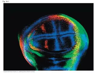
Signal Differential gene expression NUCLEUS Chromatin Chromatin modification Fig. 18-6 DNA Gene available for transcription Gene Transcription RNA Exon Primary transcript Intron RNA processing Tail mRNA in nucleus Cap Transport to cytoplasm CYTOPLASM mRNA in cytoplasm Translation Degradation of mRNA Polypeptide Protein processing Active protein Degradation of protein Transport to cellular destination Cellular function
Signal NUCLEUS Fig. 18-6a Chromatin Chromatin modification DNA Gene available for transcription Gene Transcription Exon RNA Primary transcript Intron RNA processing Tail mRNA in nucleus Cap Transport to cytoplasm CYTOPLASM
CYTOPLASM mRNA in cytoplasm Fig. 18-6b Translation Degradation of mRNA Polypeptide Protein processing Active protein Degradation of protein Transport to cellular destination Cellular function
Genes within highly packed heterochromatin are usually not expressed Chemical modifications to histones and DNA of chromatin influence both chromatin structure and gene expression Histone modification DNA methylation: genomic imprinting Epigenetic inheritance; modification In fruitfly, histone-modifying enzymes recruits a DNA methylase to silence a particular set of genes. Regulation of Chromatin Structure
Fig. 18-7 Histone tails Amino acids available for chemical modification DNA double helix (a) Histone tails protrude outward from a nucleosome Unacetylated histones Acetylated histones (b) Acetylation of histone tails promotes loose chromatin structure that permits transcription
In histone acetylation, acetyl groups are attached to positively charged lysines in histone tails This process loosens chromatin structure, thereby promoting the initiation of transcription The addition of methyl groups (methylation) can condense chromatin; The addition of phosphate groups (phosphorylation) next to a methylated amino acid can loosen chromatin Histone Modifications
Chromatin-modifying enzymes provide initial control of gene expression by making a region of DNA either more or less able to bind the transcription machinery Organization of a typical eukaryotic gene The roles of transcription factors Enhaner and specific transcription factors Regulation of Transcription Initiation
Fig. 18-8-1 Poly-A signal sequence Enhancer (distal control elements) Proximal control elements Termination region Exon Intron Exon Intron Exon DNA Upstream Downstream Promoter
Fig. 18-8-2 Poly-A signal sequence Enhancer (distal control elements) Proximal control elements Termination region Exon Intron Exon Intron Exon DNA Upstream Downstream Promoter Transcription Exon Intron Exon Intron Exon Primary RNA transcript Cleaved 3 end of primary transcript 5 Poly-A signal
Fig. 18-8-3 Poly-A signal sequence Enhancer (distal control elements) Proximal control elements Termination region Exon Intron Exon Intron Exon DNA Upstream Downstream Promoter Transcription Exon Intron Exon Intron Exon Primary RNA transcript Cleaved 3 end of primary transcript 5 RNA processing Intron RNA Poly-A signal Coding segment mRNA 3 Start codon Stop codon Poly-A tail 3 UTR 5 Cap 5 UTR
Promoter Activators Gene DNA Distal control element Enhancer TATA box Fig. 18-9-1
Promoter Activators (repressors) Gene DNA Distal control element Enhancer TATA box Fig. 18-9-2 General transcription factors DNA-bending protein Group of mediator proteins
Promoter Activators Gene DNA Distal control element Enhancer TATA box Fig. 18-9-3 ≥≥10 control element The particulat combination of control elements in an enhancer General transcription factors DNA-bending protein Group of mediator proteins RNA polymerase II RNA polymerase II Transcription initiation complex RNA synthesis
How the use of different combinations of just a few control elements can allow differential regulation of transcription in two cell types?
Enhancer Promoter Albumin gene Control elements Fig. 18-10 Crystallin gene LIVER CELL NUCLEUS LENS CELL NUCLEUS Available activators Available activators Albumin gene not expressed Albumin gene expressed Crystallin gene not expressed Crystallin gene expressed (a) Liver cell (b) Lens cell
Unlike the genes of a prokaryotic operon, each of the coordinately controlled eukaryotic genes has a promoter and control elements These genes can be scattered over different chromosomes, but each has the same combination of control elements Copies of the activators recognize specific control elements and promote simultaneous transcription of the genes Coordinately Controlled Genes in Eukaryotes
Transcription alone does not account for gene expression Regulatory mechanisms can operate at various stages after transcription Such mechanisms allow a cell to fine-tune gene expression rapidly in response to environmental changes Mechanisms of Post-Transcriptional Regulation
Exons Fig. 18-11 DNA • In alternative RNA splicing, different mRNA molecules are produced from the same primary transcript, depending on which RNA segments are treated as exons and which as introns Troponin T gene Primary RNA transcript RNA splicing or mRNA
Protein Processing and Degradation Fig. 18-12 Proteasome and ubiquitin to be recycled Ubiquitin Proteasome Ubiquitinated protein Protein to be degraded Protein fragments (peptides) Protein entering a proteasome
Only a small fraction of DNA codes for proteins, rRNA, and tRNA A significant amount of the genome may be transcribed into noncoding RNAs Noncoding RNAs regulate gene expression at two points: mRNA translation and chromatin configuration Concept 18.3: Noncoding RNAs play multiple roles in controlling gene expression
Hairpin miRNA Hydrogen bond Fig. 18-13 Dicer miRNA miRNA- protein complex 5 3 (a) Primary miRNA transcript mRNA degraded Translation blocked (b) Generation and function of miRNAs
MicroRNAs (miRNAs) are small single-stranded RNA molecules that can bind to mRNA These can degrade mRNA or block its translation The phenomenon of inhibition of gene expression by RNA molecules is called RNA interference (RNAi) RNAi is caused by small interfering RNAs (siRNAs) —ds RNA siRNAs and miRNAs are similar but form from different RNA precursors Effects on mRNAs by MicroRNAs and Small Interfering RNAs
siRNAs play a role in heterochromatin formation and can block large regions of the chromosome Small RNAs may also block transcription of specific genes Chromatin Remodeling and Silencing of Transcription by Small RNAs
During embryonic development, a fertilized egg gives rise to many different cell types Cell types are organized successively into tissues, organs, organ systems, and the whole organism Gene expression orchestrates the developmental programs of animals Concept 18.4: A program of differential gene expression leads to the different cell types in a multicellular organism
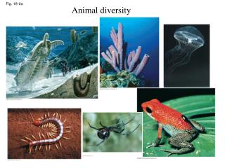
Fig. 18-14 (a) Fertilized eggs of a frog (b) Newly hatched tadpole • The transformation from zygote to adult results from cell division, cell differentiation, and morphogenesis
Fig. 18-15 Unfertilized egg cell Sperm Nucleus Fertilization Two different cytoplasmic determinants NUCLEUS Early embryo (32 cells) Zygote Signal transduction pathway Mitotic cell division Signal receptor Signal molecule (inducer) Two-celled embryo (b) Induction by nearby cells (a) Cytoplasmic determinants in the egg
Determination commits a cell to its final fate Determination precedes differentiation Cell differentiation is marked by the production of tissue-specific proteins Sequential Regulation of Gene Expression During Cellular Differentiation
Nucleus Master regulatory gene myoD Other muscle-specific genes DNA Embryonic precursor cell Fig. 18-16-1 OFF OFF
Nucleus Master regulatory gene myoD Other muscle-specific genes DNA Embryonic precursor cell Fig. 18-16-2 OFF OFF OFF mRNA MyoD protein (transcription factor) Myoblast (determined)
Nucleus Master regulatory gene myoD Other muscle-specific genes DNA Embryonic precursor cell Fig. 18-16-3 OFF OFF OFF mRNA MyoD protein (transcription factor) Myoblast (determined) mRNA mRNA mRNA mRNA Myosin, other muscle proteins, and cell cycle– blocking proteins MyoD Another transcription factor Part of a muscle fiber (fully differentiated cell)
Pattern formation is the development of a spatial organization of tissues and organs In animals, pattern formation begins with the establishment of the major axes Positional information, the molecular cues that control pattern formation, tells a cell its location relative to the body axes and to neighboring cells Pattern Formation: Setting Up the Body Plan
In Drosophila, cytoplasmic determinants in the unfertilized egg determine the axes before fertilization After fertilization, the embryo develops into a segmented larva with three larval stages The Life Cycle of Drosophila
Thorax Head Abdomen 0.5 mm Dorsal Fig. 18-17 Right BODY AXES Anterior Posterior Left Ventral (a) Adult Follicle cell Egg cell developing within ovarian follicle 1 Nucleus Egg cell Nurse cell Egg shell Unfertilized egg 2 Depleted nurse cells Fertilization Laying of egg Fertilized egg 3 Embryonic development Segmented embryo 4 0.1 mm Body segments Hatching Larval stage 5 (b) Development from egg to larva