Download
1 / 23
260 likes | 560 Views
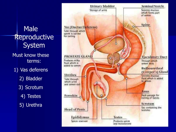
Testis, epididymis, vas deferens and spermatic cord. OBJECTIVES. OBJECTIVES. I- Primary Sex Organ: Testis. II- Reproductive Tract: Epididymis. Vas Deferens. Spermatic cord. III- Accessory Sex Glands: Seminal vesicles. Prostate gland. Bulbourethral glands.

E N D
I- Primary Sex Organ: Testis. II- Reproductive Tract: Epididymis. Vas Deferens. Spermatic cord. III- Accessory Sex Glands: Seminal vesicles. Prostate gland. Bulbourethral glands. IV- External genitalia: Penis Components of Male Reproductive System
Scrotum T L The scrotum is a cutaneous sac consisting of two layers: heavily pigmented skin and closely related dartosfascia It is divided into two compartments which are distinguished externally by a middle ridge called the raphe. Internally, the raphe connects to a muscular partition, the septum, which serves to divide the scrotum into its two areas The Left scrotum is lower than the right.
Contents: One testis and one epididymis in each compartment • Functions: • Houses & Protects the testis • Regulates testicular temperature • It has thin skin with sparse hairs and sweat glands. • The Dartos muscle lies within the superficial fascia.
The arterial supply of the scrotum : 1- Posterior scrotal branches of the perineal artery, a branch of the internal pudendal artery. 2- Anterior scrotal branches of the deep external pudendal artery, a branch of the femoral artery. 3- Cremastericartery, a branch of the inferior epigastric artery.
Scrotal veins accompany the arteries • Lymphatic vessels of the scrotum drain into the superficial inguinal lymph nodes
The nerves of the scrotum include the: • Genital branch of the genitofemoral nerve (L1, L2) supplying the anterolateral surface. • Anterior scrotal nerves, branches of the ilioinguinal nerve (L1) supplying the anterior surface. • Posterior scrotal nerves, branches of the perineal branch of the pudendal nerve (S2- S4) supplying the posterior surface. • Perineal branches of the posterior femoral cutaneous nerve (S2, S3) supplying the inferior surface.
Paired almond-shape gonads that are suspended in the scrotum by the spermatic cord 4 - 5 cm long Weigh (10.5 – 14) g Functions: Spermatogenesis. Hormone production (Androgens- testosterone). Testis sc T
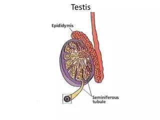
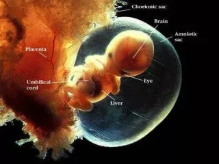
-It develops in the abdomen; descends into the scrotum before birth; -It is made up of tightly coiled tubes (SEMINIFEROUS TUBULES) where sperm are formed; -scattered between tubules are the LEYDIG CELLS(produce testosterone and other androgens).
(Leydig cells) TESTIS
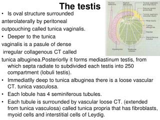
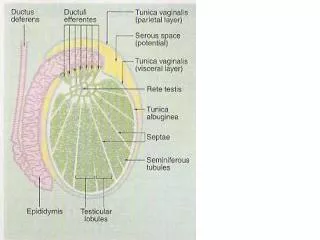
Coverings of the Testis TV Tunica Vaginalis: Peritoneal covering, formed of parietal and visceral layers. It surrounds testis & epididymis. It allows free movement of testis inside scrotum. Tunica albugenia It is a whitish fibrous capsule
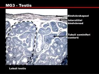
Internal Structure of Testis Fibrous septae extend from the capsule, divide the testis into a (200-300) lobules. Each lobule contains, (1-3) seminiferous tubules. Seminiferous Tubules: They are the site of the spermatogenesis. They form the bulk of testicular tissue. Rete testis: (a network of tubules) It is the Site of merging of the Seminiferous tubules. RT
Blood Supply of Testis Testicular Artery Testicular artery: It is a direct branch from the abdominal aorta. Venous drainage : (Pampiniform plexus of veins. Approximately a dozen veins which forms a network in the spermatic cord. They become larger, converge as it approached the inguinal canal and form the Testicular vein.
Right Vein drains into IVC. Left Vein drains into Left Renal Vein. Testicular Lymphatics: Follow arteries, veins end in Lumbar (par aortic) nodes From scrotum, penis, prepuce: Terminate in Superficial Inguinal nodes
Epididymis H V D A Single coiled tubule 6 M long Located on the posterior & superior margins of the testis. It is divided into: Head, Body and Tail. The Headreceives efferent ductules from testis. The Tail is continuous with Vas Deferens B T
Functions: • 1. Secretes/absorbs the nourishing fluid. • 2. Recycles damaged spermatozoa. • 3. Stores spermatozoa Up to 2 weeks to allow for maturation. • It is the site where sperms become motile and gain the ability to fertilize.
A Muscular tube 45 cm long. Carries sperms from the Epididymis to pelvic cavity. Passes through the inguinal canal It crossesthe ureter Its terminal part is dilated to form the Ampulla of the vas It joins the urethra in the prostate Vas Deferens