Download
1 / 12
140 likes | 833 Views
Regeneration of damaged intervertebral disc. Masoud Ghorbani(1),Mohammad Vasei(2*),Jafar Ai(2),Mohammadreza Nourani(1),Mahmoud Azami(2),Batool Hashemibeni(3) 1- Applied biotechnology research center, Baqiatallah university of medical sciences,Tehran,Iran

E N D
Regeneration of damaged intervertebral disc Masoud Ghorbani(1),Mohammad Vasei(2*),Jafar Ai(2),Mohammadreza Nourani(1),Mahmoud Azami(2),Batool Hashemibeni(3) 1- Applied biotechnology research center, Baqiatallah university of medical sciences,Tehran,Iran 2- Tissue Engineering and applied cell sciences department,Tehran university of medical sciencesD 3-Anatomical and molecular sciences,Medical school,Isfahan university of medical sciences Personal Photo Electronic Poster Code: A-10-650-1 MasoudGhorbani-Applied Biotechnology Research center,Baqiatallah university of medical sciences, Tehran, Iran
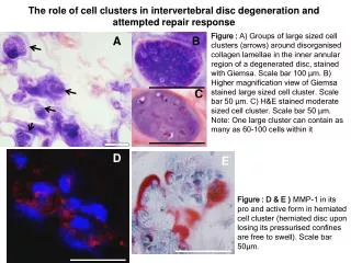
Introduction: Intervertebral disc degeneration(IVD)(Fig B,C) is recognized to be the leading cause for chronic low back pain(LBP)(Fig a). Herniation of IVD and its degeneration (Fig D) are the major reasons of low back pain, which occur because of structural damage of disc and is caused of herniation. In support forces that carry heavy military equipment, Heavy Equipment can lead to LBP or herniation. In war and conflicts, bullet and crack can damage spine and discs and lead to disability.Current methods for treatment of degeneration are artificial dsic and prothesises but there are caused of limitation of movment and are invasive.Advance Method is tissue engineering that cell,hydrogel and growth factor injecte to disc and repair it. Injectable hydrogels have various application.using of stem cell on hydrogel increase rate of repair and differentiated to cells of disc that caused of secration of extracellular matrix in degenerated disc and repair. A goal of this study was Synthesis hydrogel similar to extracellular matrix of disc named injectable compound hydrogel and with NP and BMSc injected to degenerated disc of animal rabbit and suvey effects of it in regeneration of degenerated disc. Fig A Fig B Fig C Fig D MasoudGhorbani-Applied Biotechnology Research center,Baqiatallah university of medical sciences, Tehran, Iran
Method: Invitro study: compound hydrogel synthesis with chitosan, B glycerol Phosphate, collagen, gelatin, hyaluronic acid, chondroitin 6 sulfate and fibroin silk. Rheology(Time Gelation, Temperature gelation), MTT(Viability), trypan blue(Cell number), SEM(Porosity) and RT-PCR(expression of Sox9,Type I and II collagen) used for evaluation of hydrogel. Invivo study: Discs 3-6 in animal model degenerated using by needle and injection of papain . Bone marrow mesenchymal stem cells (BMSc) extracted from rabbit,cultured,encapsulated into hydrogel and injected to degenerated disc. Finally, effects repair of cells and hydrogel evaluated by MRI(High disc,intensity), histology(border line,allinedcollagen,cells of dic),IFC(Type II collagen). MasoudGhorbani-Applied Biotechnology Research center,Baqiatallah university of medical sciences, Tehran, Iran
Time sweep: result shows the changes of G′ and G″ versus time measured at 37 °C. The gelationpoint is the timewhenstorage and lossmodulus (G′ and G″) become equal. Thus the gelation time of NP hydrogel was about 30 min.(Fig. 1). MTT: OD of wells in which cells cultured with hydrogel, increased insignificantly. It means that the survival of NP cells cultured beside NP hydrogel at day 0–21 increased, and this increasewas statistically significant(Fig2). Trypan Blue: Results of cell count showed an increased insignificantly. It means that the Cell number and viability of NP cells in sided compound hydrogel at day 0–21 increased and this increase was statistically significant .The viability of NP cells in NP hydrogel compared to the control group was significant and too (Fig3). Result: Invitro study: Fig3:Comparison percent of alive NP cells in NP hydrogel and control groups (*P < 0.05).) Fig1: Changes of storage and loss modulus (G′ and G″) versus time for the gel solution in 37oC for estimation of gelation time. Fig2:Result of MTT assay to investigate survival of NP cells cultured in NP hydrogel and control groups at day 0 to 21 days (*P < 0.05). MasoudGhorbani-Applied Biotechnology Research center,Baqiatallah university of medical sciences, Tehran, Iran
SEM:The surfaces of the NP hydrogels (Fig 4) contain many pores, which vary in size, although mostwere smaller than 200min diameter. This hydrogel had high porosity that is suitable for tissue engineering of IVD. RT-PCR:All groups showed significant increase in type 1 and 2collagen and aggrecan and sox9 in comparison to their control (Fig3). At 2 and 3 months, there were a significant difference for sox9 and aggrecan in H-N group in compared to the other groups. At months 2, there was a significant difference for type 2 collagen in H-N and at months 3 in H-S group. At months 3, there was significant difference for type 1 collagen in D and H-S groups. At month 2, H-NP had the significant highest expression type 2 collagen, Sox9 and aggrecan .The D group showed the highest expression of type 1 collagen (Fig3 b, d, and f). At 3 months, H-NP showed the significant highest expression Sox9 and aggrecan .On other hand; H-S indicated the significant highest expression type 2 collagen. The D group showed the significant highest type 1 collagen. Result: Invitro study: Fig5: expression of coll1, 2, Sox9 normalized with GAPDH in degeneration (D), BMS-hydrogel(H-S) and NP-hydrogel(H-N)groups compared with control group(P<0.05 was significant). Fig4: SEM micrograph of NP hydrogel surface. MasoudGhorbani-Applied Biotechnology Research center,Baqiatallah university of medical sciences, Tehran, Iran
H&E staining: Results showed the presence of aligned fibroblast-like cells (AF cells) in the AF region of neodisc and a large amount of round-shaped chondrocyte-like cells (NP or NC cells), in the NP region of regenerated tissues in H-S and H-N groups .Generally, H&E staining of D group displayed the lower amount of spindle-shaped fibroblast-like cells and lower amount of chondrocyte-like cells (Fig6).H&E staining (Fig6) and IFC(Fig7) revealed a well-organized arrangement of collagen I fibers, which were arranged in parallel pattern similar to those of native disc, in H-S and H-NP groups In contrast, in D group ,collagen I fibers presented irregular and discontinuous patterns with no alignments in some parts of the specimens. Result: Invivo study: Fig6: Histological sections of the native disc, degenerated disc tissue at 2 and 3 months post-injection: Native disc, degeneration disc in 2 and 3 months (D2, D3), hydrogel/stem cells in 2 and 3 months (H-S2 and H-S3) and hydrogel/NP cells in 2 and 3 months (H-N2 and H-N3) groups. In the experimental groups, staining (black arrows) showing the presence of round chondrocyte-like cells (NP and NC cells) in the AF and NP zone, aligned between collagen fibers. PC (or yellow arrows) in all the groups showing plump cells (degenerating NP cells) in Transition zoon (TZ).Red arrows showing transition zoon in the all of groups that were sharp in H-S2,H-S3,H-N2 and H-N3 groups and not sharp in D2 and D3 groups.
IFC: IFC revealed a well-organized arrangement of collagen I fibers, which were arranged in parallel pattern similar to those of native disc, in H-S and H-NP groups In contrast, in D group ,collagen I fibers presented irregular and discontinuous patterns with no alignments in some parts of the specimens. According to HE/IFC staining, sharp lines(aligned collagen fibers) in AF zoon were more and border line (between AF and NP area ) was sharp in NP-Hydrogel group in comparison to others group while, such lines not observed in D group(Fig 7) . Fig 7 : IFC images of native disc ,degeneration in 2months post degeneration(D2),degeneration in 3 months post degeneration(D3), BMS-hydrogel in 2 months(H-S2),BMS-hydrogel in 3 months(H-S3),NP-hydrogel in 2 months(H-NP2) and NP-hydrogel in 3months(H-NP3) post degeneration
MRI: MRI results indicated observable morphological changes in all groups. No evidence of disc regeneration showed in all groups in 1 month but All two treatment groups NP-hydrogel (H-N) and hydrogel-BMSc (H-S) in 2,3 months demonstrated more qualitative evidence of disc regeneration than the degeneration (punctured) group(D), as shown in Fig. 1. The signal intensity and high of disc was more in H-N and was lesser in D. MRI parameters in H-N group was most closely resembled the control (c) and before injection groups (Fig 8) . Fig 8: MRI results:T2 images of spine: normal disc(a),degeneration disc(b),2 months post injection(c),3months post injection(d).Control(C),Degeneration(D),Hydrogel-BMS(H-S) and Hydrogel-NP(H-N) groups MasoudGhorbani-Applied Biotechnology Research center,Baqiatallah university of medical sciences, Tehran, Iran
Disscusion: Our thermoresponsive hydrogel is a biocompatible chitosan based hydrogel that can be used in engineering of some tissues with injection into tissue especially for IVD regeneration .In rheometry tests we observed gelation time was about 30 min. MTT and trypan blue results showed our thermo responsive hydrogel was cyto compatible and safe complex biomaterial that maintains viability and proliferation of NP cells and can use for many tissues. Yung-Hsin Cheng et al .cultured rabbit NP cells in gelatin/ the thermosensitive chitosan/b-glycerol phosphate . Injectable hydrogels are an important category of biomaterials in tissue engineering .Softness, flexibility, injectability, easily casting into different shapes and three dimensional structures of hydrogels which mimic the extra cellular matrix of natural tissues make them very useful in this field. evaluations revealed production of ECM in all groups . In NP-Hydrogel group, sharp lines(aligned collagen fibers) in AF zoon were more , NP cells was abundantly and border line (between AF and NP area ) was sharp in in comparison to others group. In BMSc-hydrogel group, Sharp lines were more, NP cells were lesser and border line was sharper in comparison to degeneration group. These results showed the NP-hydrogel group was anatomically and histologically more similar to native disc than those of the other groups. In IFC staining, collagen type 2 was strongly positive in NP-hydrogel group . RT-PCR confirmed histology results. The significant increase type 1,2collagens, aggrecan and Sox9 was observed for NP-hydrogel group as showed by Real-Time PCR .It may be because of 1.positive effect transplanted NP cells in production of ECM 2.the presence of transplanted or remaining NP cells after degeneration 3. Create an ideal environment for proliferation and production of cells using by injected hydrogel. Increase of signal intensity and high of disc was sign of proliferation of NP cells, produce of ECM and hydration in NP-hydrogel group. In contrast, decrease of these parameters in degeneration group may be because of dehydration, lack of NP cells and reduce speed of self-regeneration. MasoudGhorbani-Applied Biotechnology Research center,Baqiatallah university of medical sciences, Tehran, Iran
Conclusion: In this study, an injectable hydrogel was prepared and utilized for disc regeneration in a rabbit model. In our study, the NP cell-hydrogel well promoted NP tissue regeneration. In vivo and in vitro experimental results related to NP-hydrogel group were approximately close to native disc. We can conclude that the injection of hydrogel-NP cells after degeneration induce regeneration associated with production of ECM in the degenerated disc, resulting in improvement of the disc structure 3 months after degeneration. We conclude that NP cell can contribute as an appropriate cell source to regeneration of NP tissue. Natural compound hydrogel synthesized in pervious invitro study that was biocompatible, injectable with high porosity and properties structure similar to ECM of IVD Although seeding of NP cells on injectable hydrogel may provide an important insight into disc regeneration, but further in vivo experiments with long term follow-up on larger animal models are required to assess the clinical validity of this hydrogel for disc tissue engineering and compare role of cells and hydrogel separately in further groups. MasoudGhorbani-Applied Biotechnology Research center,Baqiatallah university of medical sciences, Tehran, Iran
References [1] G. Waddell, Low back pain, A twentieth century health care enigma, Spine 21 (1966) 20–25. [2] N. Boss, R. Rieder, V. Schade, K.F. Spartt, N. Semmer, Aebim, The diagnostic accuracy of magnetic resonance imaging, work perception, and psychosocial factors in identifying symptomatic descherniations, Spine 20 (1995) 2613–2625. [3] S. Sobajima, G. Vadala, A. Shimer, J.S. Kim, L.G. Gilbertson, J.D. Kang, Feasibility of a stem cell therapy for intervertebral disc degeneration, Spine J. 8 (2008) 888–896. [4] TabatoY, Recent progress in tissue engineering, Drug Discov. Today 6 (2001) 483–487. [5] K. Tsuchiya, G. Chen, T. Ushida, T. Matsuno, T. Tateishi, The effect of coculture of chondrocytes with mesenchymal stem cells on their cartilaginous phenotype in vitro, Mater. Sci. Eng. 24 (2004) 391–396. [6] A. Atala, Tissue engineering and artificial organs, J. Endourol. 14 (2000) 49–57. [7] G. Vunjak-Novakovic, R.I. Freshney, Culture of Cells for Tissue Engineering, Wiley Online Library, 2006. [8] T. Haringham, S. Tew, A. Murdoch, Tissue engineering: chondrocyes and cartilage, Arthritis Res. 4 (2002) 63–68. [9] C. Hoemann, J. DSun, A. Legare, M.D. Mckee, M.D. Buschmann, Tissue engineering of cartilage using an injectable and adhesive chitosan-based cell-delivery vehicle, Osteoarthr. Cartil. 13 (2005) 318–329. [10] D. Macaya, M. Spector, Injectable hydrogel materials for spinal cord regeneration, Biomed. Mater. 7 (2012) 1–22. [11] R. Ahmadi, J.D. deBruijn, Biocompatibility and gelation of chitosan–glycerophosphate hydrogels, Biomed. Mater. Res. 86 (2008) 824–831. [12] J. Yan, L. Yang, G. Wang, Y. Xiao, B. Zhang, N. Qi, Biocompatibility evaluation of chitosan- based injectable hydrogels for the culturing mice mesenchymal stem cells in vitro, Biomater. Appl. 24 (2010) 625–636. [13] M. Barikani, E. Oliaei, H. Seddiqi, H. Honarkar, Preparation and application of chitin and its derivatives: a review, Iran. Polym. J. 23 (2014) 307–326. [14] A. Laiji, A. Sohrabi, D.S. Hungerford, C.G. Frondoza, Chitosan supports the expression of extracellular matrix proteins in human osteoblasts and CSndrocytes, J. Biomed. Mater. 51 (2000) 586–595. [15] J.Y. Lee, et al., Enhanced bone formation by controlled growth factor delivery from chitosan-based biomaterials, J. Control. Release 78 (2002) 187–197. [16] A. Chenite, C. Chaput, D. Wang, C. Combes, M.D. Buschmann, C.D. Hoemann, J.C. Leroux, B.L. Atkinson, F. Binette, A. Selmani, Novel injectable neutral solutions of chitosan form biodegradable gels in situ, Biomaterials 21 (2000) 2155–2161. MasoudGhorbani-Applied Biotechnology Research center,Baqiatallah university of medical sciences, Tehran, Iran
Final Questions • What is already known about the subject? • Tissue engineering using by injectable hydrogel and stem cells can use for regeneration of degenerated intervertebral dis. • What could be added to the current knowledge by this study? • We conclude that NP cell can contribute as an appropriate cell source to regeneration of NP tissue. NP cells on injectable hydrogel may provide an important insight into disc regeneration MasoudGhorbani-Applied Biotechnology Research center,Baqiatallah university of medical sciences, Tehran, Iran